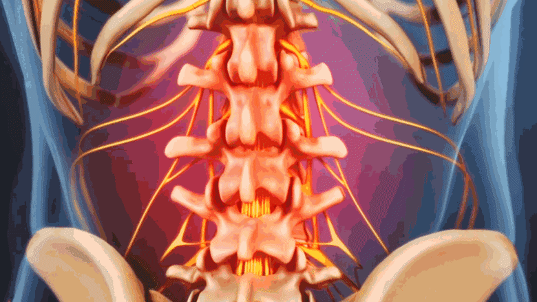
Discography or discogram for back pain and sciatica involves injecting a contrast agent into a disc suspected to be the cause of pain. The contrast agent increases the pressure within the disc and provokes its painful symptoms. Through continuous medical imaging, such as x-rays or computed tomography (CT) scans, taken at the same time, the contrast agent captures the disc’s anatomical structure.1Peh W. Provocative discography: Current status. Biomed Imaging Interv J. 2005;1(1):e2. doi:10.2349/biij.1.1.e2 Available from: https://www.ncbi.nlm.nih.gov/pmc/articles/PMC3097593/
The goal of a discogram test is to:
- Evaluate the symptoms caused by provoking the disc and analyze if they match with the patient’s everyday, familiar symptoms1Peh W. Provocative discography: Current status. Biomed Imaging Interv J. 2005;1(1):e2. doi:10.2349/biij.1.1.e2 Available from: https://www.ncbi.nlm.nih.gov/pmc/articles/PMC3097593/
- Assess the extent of damage to the disc, which may help provide the rationale for surgical treatment of the involved spinal segment1Peh W. Provocative discography: Current status. Biomed Imaging Interv J. 2005;1(1):e2. doi:10.2349/biij.1.1.e2 Available from: https://www.ncbi.nlm.nih.gov/pmc/articles/PMC3097593/
The test is mostly conducted by physiatrists, anesthesiologists, and radiologists with special training in diagnosing spinal pain through minimally invasive procedures. It is important to educate patients that this test will cause some degree of discomfort or pain but will not be unbearable. Patients are advised to interpret every stimulation that they may feel throughout the procedure to help make the diagnosis as accurate as possible. When pain that is closest to the familiar pain is experienced, the test is considered positive.
In This Article:
- Lumbar Discography for Back Pain Diagnosis
- Lumbar Discogram Procedure
- Potential Side Effects and Risks of Discogram
Preparation Before Lumbar Discogram
A physical exam identifies pain patterns and rules out red-flag symptoms like spinal cord compression.
A thorough physical exam and medical history are important steps in preparing for a discogram.
- The physical exam helps the physician understand the patient’s pain pattern(s) stemming from the suspected disc and rule out any red flag symptoms, such as numbness in the groin area or severe numbness and weakness in the leg(s), which may indicate underlying medical emergencies and require immediate treatment.
- A medical history can provide an understanding of the patient’s past treatments and medication, which may help the physician determine if surgical intervention is advisable for the disc-related problem.
The medical history also includes a review of past diagnostic test results, such as x-rays, CT scans, and/or magnetic resonance imaging (MRI) scans, which are carefully reviewed and used in conjunction with discogram results for improved diagnosis and interpretation of disc issues.
Patients are advised to fast for 6 to 8 hours before the test.1Peh W. Provocative discography: Current status. Biomed Imaging Interv J. 2005;1(1):e2. doi:10.2349/biij.1.1.e2 Available from: https://www.ncbi.nlm.nih.gov/pmc/articles/PMC3097593/
A discogram is performed in a hospital on an outpatient basis. The procedure takes about an hour and patients usually return home the same day.
- The discogram procedure is performed while the patient lies on his or her stomach or side.
- An intravenous line of medications containing antibiotics is started to help prevent infection of the disc and spinal tissues.1Peh W. Provocative discography: Current status. Biomed Imaging Interv J. 2005;1(1):e2. doi:10.2349/biij.1.1.e2 Available from: https://www.ncbi.nlm.nih.gov/pmc/articles/PMC3097593/
- Sedation is usually avoided as it may interfere with the pain provocation response of the patient. If sedation is given, a mild dose may be administered.1Peh W. Provocative discography: Current status. Biomed Imaging Interv J. 2005;1(1):e2. doi:10.2349/biij.1.1.e2 Available from: https://www.ncbi.nlm.nih.gov/pmc/articles/PMC3097593/
- Local anesthesia is used to anesthetize a core of tissue from the skin to the disc’s surface.
- The skin over the injection site is cleaned and a sterile needle is guided toward the disc using live x-ray or CT scan.
- When the needle touches the outer surface (annulus fibrosus) of the disc, a distinctly smaller needle is advanced through the first needle (double-needle technique) and progressed into the core of the disc (nucleus pulposus).
- The disc is then pressurized by injecting a contrast agent into its core through the smaller needle.
- At this point, the patient’s response is documented.
- Pain that is different from what is familiar and felt every day indicates that the pressurized disc is not symptomatic
- Pain that is similar to what is familiar and felt every day indicates that the pressurized disc is symptomatic
- After the disc is pressurized and the patient’s responses are acknowledged, pictures of the disc are taken with the fluoroscopic unit and the needles are removed.
The procedure may be repeated on more than one disc. Some physicians use a single-needle technique, where only one needle is advanced from the skin’s surface to meet the core of the disc.1Peh W. Provocative discography: Current status. Biomed Imaging Interv J. 2005;1(1):e2. doi:10.2349/biij.1.1.e2 Available from: https://www.ncbi.nlm.nih.gov/pmc/articles/PMC3097593/
Performing a discogram in the lumbar spine needs precision, training, technique, and experience. It’s important to note that interpreting a patient’s pain response is subjective; if the procedure is performed by an inexperienced physician, it can result in misinterpretation of the result and potentially unnecessary surgery.
Recovery and Transient Side Effects After Lumbar Discogram
Back pain might persist for a few days following a discogram.
The patient is carefully examined for vital signs and any evidence of bleeding or blood clots for about 30 minutes after the discogram and discharged if no concerns are present.2Derby R, Lee SH, Chen Y. Discograms: Cervical, thoracic, and lumbar. Techniques in Regional Anesthesia and Pain Management. 2005;9(2):97-105. doi.org/10.1053/j.trap.2005.05.009
Lingering back pain may be present for a few days after the procedure.2Derby R, Lee SH, Chen Y. Discograms: Cervical, thoracic, and lumbar. Techniques in Regional Anesthesia and Pain Management. 2005;9(2):97-105. doi.org/10.1053/j.trap.2005.05.009 The injection site may be treated with over-the-counter pain-relieving medications and/or an ice pack. A flare-up of the usual symptoms stemming from the disc may occur for 2-7 days; rarely, the flare-up may continue for a longer period of time.2Derby R, Lee SH, Chen Y. Discograms: Cervical, thoracic, and lumbar. Techniques in Regional Anesthesia and Pain Management. 2005;9(2):97-105. doi.org/10.1053/j.trap.2005.05.009
When administered by a skilled professional, discogram injections are generally considered safe and may be a useful presurgical diagnostic tool for suitable patients with chronic back pain. In rare cases, serious adverse reactions may occur, which can largely be prevented by using standard precautions.
- 1 Peh W. Provocative discography: Current status. Biomed Imaging Interv J. 2005;1(1):e2. doi:10.2349/biij.1.1.e2 Available from: https://www.ncbi.nlm.nih.gov/pmc/articles/PMC3097593/
- 2 Derby R, Lee SH, Chen Y. Discograms: Cervical, thoracic, and lumbar. Techniques in Regional Anesthesia and Pain Management. 2005;9(2):97-105. doi.org/10.1053/j.trap.2005.05.009