
Subscribe to our newsletter and get your FREE guide,
Natural Back Pain Relief: 16 Choices for Lasting Comfort.
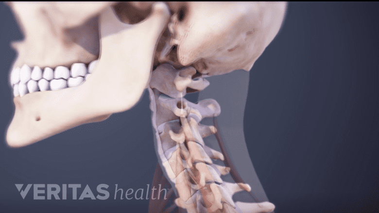
Neck muscles help support the cervical spine and contribute to movements of the head, neck, upper back, and shoulders.
In This Article:
- Cervical Spine Anatomy
- Cervical Vertebrae
- Neck Muscles and Other Soft Tissues
- Cervical Discs
- Cervical Spinal Nerves
- Spinal Cord Anatomy in the Neck
- Cervical Spine Anatomy Video
Important Muscles of the Neck
Here are some of the key muscles attached to the cervical spine:
- Levator scapulae. The levator scapulae muscle is attached at the top four cervical vertebrae (C1 to C4) and runs down the side of the neck to attach at the top of the shoulder blade (scapula). This muscle helps with lifting the shoulder blade, bending the neck to the side, and rotating the head.
- Sternocleidomastoid (SCM). The SCM muscle is attached to a small bone behind the ear (called the mastoid process) and travels down the front of the neck to attach at both the sternum and collarbone. Depending on whether one or both SCM muscles (one on each side of the neck) are contracted, the head can be rotated to the side or the chin tilted upward. It is a large muscle that also helps protect some fragile structures, such as the carotid artery.
- Trapezius. The trapezius muscle is a large surface muscle that spans from the base of the skull down the cervical spine and into the lower thoracic spine (mid back), as well as out to the shoulder blade. The two trapezius muscles together form a kite shape. The trapezius muscle can be involved in extending the head upward or neck backward, rotating/turning the head, or lifting the shoulder blade.
- Erector spinae. Numerous muscles comprise the erector spinae muscles throughout the spine. In the cervical spine, the erector spinae muscles play key roles in supporting posture, rotating the neck, and extending the neck backward.
- Deep cervical flexors. The muscle group is comprised of the longus capitus and longus colli muscles, which run down the front of the cervical spine. The deep cervical flexor muscles are involved in flexing the neck forward as well as stabilizing the cervical spine.
- Suboccipitals. Comprised of 4 pairs of small muscles, the suboccipital muscles connect the top of the cervical spine with the base of the skull. The suboccipitals are important for head extension and rotation.
See Forward Head Posture’s Effect on Neck Muscles
There are numerous other muscles connected to the neck, which all work in concert with tendons (connect muscles to bones) and ligaments (connect bones to bones). If a neck muscle is strained, it can become painful, tight, and possibly lead to a stiff neck.
advertisement
Cervical Spine Ligaments
Ligaments connect one bone to the other.
Ligaments are durable soft tissues that connect bones together. There are many ligaments in the neck, but 3 types are important for helping to stabilize the entire spine:
- Anterior longitudinal ligament (ALL). This ligament starts at the base of the skull (occiput) and goes down the front of the vertebral bodies and intervertebral discs. When the neck is extended backwards, the ALL is the key ligament to resist and become stretched during that movement.
- Posterior longitudinal ligament (PLL). The PLL starts at C2 and goes down the back of the vertebral bodies and intervertebral discs. When the neck is flexed forward, this ligament stretches and resists that motion. The PLL is located inside the spinal canal in front of the spinal cord.
- Ligamentum flava. The ligamentum flava are short, paired ligaments that connect the vertebral arches of adjacent vertebrae, helping to cover the spinal cord from behind. Starting at C2-C3, each ligamentum flavum connects a lamina to the lamina below. Over time, the ligament flava thicken and in some cases contribute to compressing a spinal nerve root or the spinal cord.

There are numerous other ligaments that play a part in holding neck bones together, which is crucial for other neck functions to occur, such as facilitating blood flow through the cervical spine.
Blood Supply of the Cervical Spine
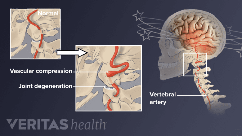
Compression of the vertebral artery in the neck can cause dizziness and/or headache.
The vertebral artery is critical to supplying blood from the heart to the brain and spinal cord. A right and left vertebral artery travel up the cervical spine symmetrically. The vertebral artery typically follows this general path:
- Branches from the subclavian artery above the heart and goes into a bony hole (foramen) in C6’s transverse process.
- Travels straight up the cervical spine through the foramina from C6 to C2.
- Takes more of a winding route when traveling from C2 (axis) to and through the foramen of C1 (atlas).
- Travels from C1 and goes beneath the posterior atlanto-occipital membrane (which stretches from the top of C1 to the base of the skull) to enter the spinal cord.
advertisement
Once inside the spinal cord, the right and left vertebral arteries join in the basilar artery to supply the brainstem. There are also many arteries that branch off the various levels of the vertebral arteries to send blood to the spinal cord, bones, joints, and other areas.
If a vertebral artery starts to become compressed due to spinal degeneration or injury, it can cause neck pain, headache, and/or possibly dizziness..

advertisement