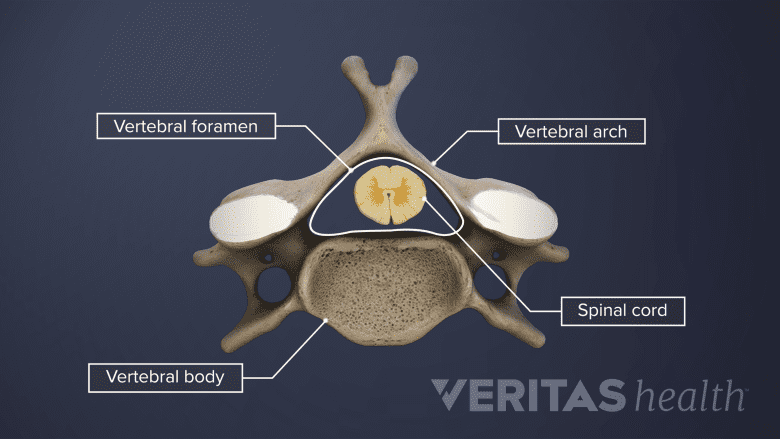
The spinal cord is a bundle of nerves that carry electrical signals between the brain and the rest of the body. One of the most important jobs of the cervical spine is to protect the spinal cord as it travels through the neck to innervate the rest of the body.
In This Article:
- Cervical Spine Anatomy
- Cervical Vertebrae
- Neck Muscles and Other Soft Tissues
- Cervical Discs
- Cervical Spinal Nerves
- Spinal Cord Anatomy in the Neck
- Cervical Spine Anatomy Video
Internal Anatomy of the Spinal Cord

The spinal cord is housed in the spinal canal and protected by the bones and ligaments of the spine.
When viewed as a cross-section from above, the spinal cord consists of a butterfly-shaped (or thick H-shaped) region of gray matter that sits in the middle of the white matter.
The gray matter, which is primarily composed of nerve cell bodies, has two regions on each side (or butterfly wing) within the cervical spine’s region of the spinal cord:
- Posterior (dorsal) horn. This back section of the gray matter region connects with the posterior nerve root and receives sensory signals, such as for pain, temperature, and touch.
- Anterior (ventral) horn. This front section of the gray matter region connects with the anterior nerve root and sends motor signals to control muscles, such as in the neck, shoulder, arm, hand, or elsewhere.
White matter consists of axons covered in myelin (which are comprised of proteins and lipids that help protect the axons and facilitate the transmission of nerve signals). Collections of axons are called tracts. Some spinal tracts carry sensory signals up toward the brain, whereas others carry motor signals down toward muscles to control the body.
Watch Spinal Cord and Spinal Nerves: What is the Difference? Animation
In the cervical spine, the anterior horns of the gray matter are enlarged at spinal levels C4 through C8 compared to levels above and below.1Darby SA. General anatomy of the spinal cord. In: Cramer GC, Darby SA, Clinical Anatomy of the Spine, Spinal Cord, and Ans, 3rd Edition. St. Louis, MO: Elsevier; 2014: 122.
Protective Layers of the Spinal Cord

The spinal meninges help prevent the spinal cord from direct contact with the bony cervical spine.
Within the neck region, the spinal meninges help prevent the spinal cord from direct contact with the bony cervical spine. The three layers of the spinal meninges include:
- Dura mater. This tough outermost layer is constructed of dense fibrous tissue. The dura mater is the only layer of the spinal meninges that can feel pain.
- Arachnoid mater. This middle layer is comprised of elastic tissues and collagen in a spider web-like network. The arachnoid mater is named after this unique web-like structure.
- Pia mater. This innermost layer attaches to and closely lines the spinal cord and brain. Of the three meningeal layers, the pia mater is the slimmest and most delicate.
See When Neck Stiffness May Mean Meningitis
The cerebrospinal fluid, which is created within the brain, runs beneath the arachnoid mater in the subarachnoid space and above the pia mater.
Signs and Symptoms of Spinal Cord Compression

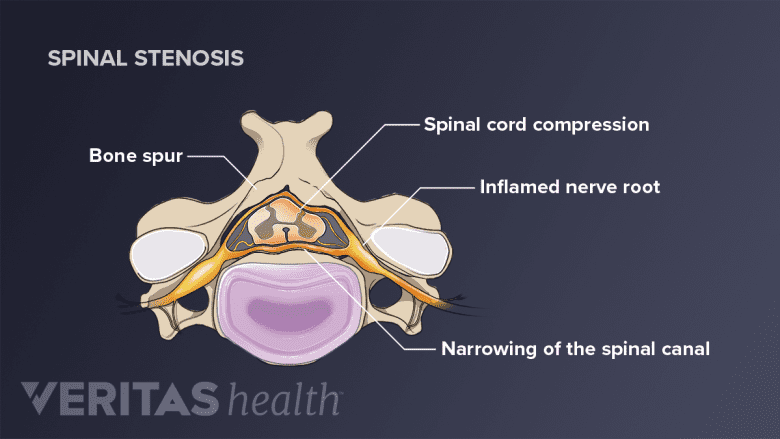
Compression of the spinal cord is a medical emergency and requires prompt treatment.
If the spinal cord becomes compressed, any number of functions involved at or below the level of compression may be affected. Usually the problems created are bilateral, that is, on both sides of the body. Some potential signs and symptoms of spinal cord compression in the neck could include one or more of the following:
- Weakness or reduced coordination. A few examples include changes in how a person walks (gait) or reduced fine motor skills in the hands.
Numbness or tingling. A pins-and-needles tingling and/or reduced ability to sense touch may be experienced in one or more regions of the body, such as the arms or legs.
Pain. If pain is present, it can range anywhere from a mild neck ache to a sharp or burning pain. For some people, bending the head forward may cause electric-like shooting pains into the arms and legs.
- Bowel and/or bladder dysfunction. Problems with controlling various bodily functions, such as the bladder and/or bowels, may develop.
There are many other possible signs and symptoms of spinal cord compression. Any neurological deficits—such as weakness, numbness, or reduced coordination—require immediate medical evaluation.
Cervical Spinal Cord Injury

Injury to the cervical spinal cord can cause temporary or permanent damage to the motor functions in the arms, torso, or legs.
Sometimes an accident or collision can lead to a more serious spinal cord injury beyond the more typical inflammation or compression associated with degenerative changes. Spinal cord injuries are typically classified by the spinal nerve level at which function is lost or impaired.
For example, a C6 spinal cord injury would result in reduced or lost function of the C6 nerves and all of the nerves below. A person with a C6 spinal cord injury would be able to breathe and move the head and shoulders well, but there would be struggles with moving the arms and likely no ability to move the trunk or legs. Sensations beneath the shoulders would also likely be impaired or lost.
- 1 Darby SA. General anatomy of the spinal cord. In: Cramer GC, Darby SA, Clinical Anatomy of the Spine, Spinal Cord, and Ans, 3rd Edition. St. Louis, MO: Elsevier; 2014: 122.

