The coccyx serves as a vital insertion site for multiple soft tissues, such as ligaments and tendons. These structures play crucial roles in supporting the pelvis, facilitating movement, and ensuring the proper functioning of the pelvic floor.1Foye PM. Coccydynia: Tailbone Pain. Phys Med Rehabil Clin N Am. 2017;28(3):539-549. doi:10.1016/j.pmr.2017.03.006
In This Article:
- Anatomy of the Coccyx (Tailbone)
- Soft Tissues and Essential Functions of the Coccyx
- Coccydynia (Tailbone Pain) Video
The soft tissues that attach to the coccyx help to maintain and preserve the ring-like structure of the pelvis; a healthy coccyx provides stability to the pelvis’ ring-like structure.
The coccyx also provides stability and support to the pelvis and helps with several movements and activities that involve the spine and legs to work together.
Coccyx Tendon Insertions

Tendons of muscles that insert into the coccyx help stabilize the pelvic floor and control spinal movements.
Tendons of essential muscles that insert into the front (anterior side) and back (posterior side) of the coccyx include1Foye PM. Coccydynia: Tailbone Pain. Phys Med Rehabil Clin N Am. 2017;28(3):539-549. doi:10.1016/j.pmr.2017.03.006,2Mabrouk A, Alloush A, Foye P. Coccyx Pain. [Updated 2023 May 1]. In: StatPearls [Internet]. Treasure Island (FL): StatPearls Publishing; 2023 Jan-. Available from: https://www.ncbi.nlm.nih.gov/books/NBK563139/:
- Levator ani: Broad, thin muscles on either side of the pelvis attach to the coccyx's anterior (front) surface and help support the pelvic organs.2Mabrouk A, Alloush A, Foye P. Coccyx Pain. [Updated 2023 May 1]. In: StatPearls [Internet]. Treasure Island (FL): StatPearls Publishing; 2023 Jan-. Available from: https://www.ncbi.nlm.nih.gov/books/NBK563139/ The levator ani is comprised of smaller muscles including:
- Iliococcygeus: A thin sheet of muscle that originates from the ischial spine and helps support the pelvic organs2Mabrouk A, Alloush A, Foye P. Coccyx Pain. [Updated 2023 May 1]. In: StatPearls [Internet]. Treasure Island (FL): StatPearls Publishing; 2023 Jan-. Available from: https://www.ncbi.nlm.nih.gov/books/NBK563139/
- Pubococcygeus: A thin sheet of muscle that extends from the front of the pelvis to the coccyx, forming a hammock-like structure to support the bladder, uterus (in females), prostate (in males), and rectum2Mabrouk A, Alloush A, Foye P. Coccyx Pain. [Updated 2023 May 1]. In: StatPearls [Internet]. Treasure Island (FL): StatPearls Publishing; 2023 Jan-. Available from: https://www.ncbi.nlm.nih.gov/books/NBK563139/
- Puborectalis: A small muscle that originates from the pubic bone in front of the pelvis and extends to the back of the rectum, playing a critical role in maintaining continence and facilitating bowel movements2Mabrouk A, Alloush A, Foye P. Coccyx Pain. [Updated 2023 May 1]. In: StatPearls [Internet]. Treasure Island (FL): StatPearls Publishing; 2023 Jan-. Available from: https://www.ncbi.nlm.nih.gov/books/NBK563139/
- Coccygeus: A small muscle that extends from the front of the coccyx to the lower sacrum and assists in supporting the pelvic floor2Mabrouk A, Alloush A, Foye P. Coccyx Pain. [Updated 2023 May 1]. In: StatPearls [Internet]. Treasure Island (FL): StatPearls Publishing; 2023 Jan-. Available from: https://www.ncbi.nlm.nih.gov/books/NBK563139/
- Sphincter ani externus: A circular muscle that surrounds the anus and attaches to the tip of the coccyx and aids in the control of bowel movements2Mabrouk A, Alloush A, Foye P. Coccyx Pain. [Updated 2023 May 1]. In: StatPearls [Internet]. Treasure Island (FL): StatPearls Publishing; 2023 Jan-. Available from: https://www.ncbi.nlm.nih.gov/books/NBK563139/
- Gluteus maximus: The largest muscle in the human body, responsible for hip and thigh movements and the only muscle attached to the posterior (back) surface of the coccyx2Mabrouk A, Alloush A, Foye P. Coccyx Pain. [Updated 2023 May 1]. In: StatPearls [Internet]. Treasure Island (FL): StatPearls Publishing; 2023 Jan-. Available from: https://www.ncbi.nlm.nih.gov/books/NBK563139/
These muscles play a crucial role in stabilizing the pelvic floor and are responsible for many bodily functions, including urination, defecation, and childbirth. Their tendinous attachments are mostly made up of collagen and are strong, flexible, and resistant to damage, similar to a fiber optic cable or a rope.
Coccyx Ligament Attachments

Injury to the coccygeal ligaments may cause tailbone pain.
Important ligaments that attach to the coccyx include:
- Anterior and posterior sacrococcygeal ligaments: The primary ligaments that help stabilize the sacrococcygeal joint between the sacrum and the coccyx. The sacrococcygeal ligaments also assist in shock absorption during walking or running.1Foye PM. Coccydynia: Tailbone Pain. Phys Med Rehabil Clin N Am. 2017;28(3):539-549. doi:10.1016/j.pmr.2017.03.006
- Lateral sacrococcygeal ligament: These ligaments are located on the side of the coccyx and have a similar function to the sacrococcygeal ligament by ensuring the coccyx stays in proper alignment with the sacrum.1Foye PM. Coccydynia: Tailbone Pain. Phys Med Rehabil Clin N Am. 2017;28(3):539-549. doi:10.1016/j.pmr.2017.03.006
- Anococcygeal ligament: The ligament that connects the tip of the coccyx to the anal hiatus (the opening in the pelvic floor through which the anal canal passes). This ligament aids in defecation and provides structural support to the rectal muscles.1Foye PM. Coccydynia: Tailbone Pain. Phys Med Rehabil Clin N Am. 2017;28(3):539-549. doi:10.1016/j.pmr.2017.03.006
- Sacrotuberous ligaments: These ligaments are attached to the coccyx on both sides and extend from the sacrum to the ischial tuberosity, a bony prominence located in the pelvis commonly known as the “sitting bone.” Along with the sacrospinous ligaments, they prevent the sacrum from tilting backward.1Foye PM. Coccydynia: Tailbone Pain. Phys Med Rehabil Clin N Am. 2017;28(3):539-549. doi:10.1016/j.pmr.2017.03.006
- Sacrospinous ligaments: These ligaments are attached to the coccyx on both sides and extend from the outer edge of the sacrum to the ischial spine, one of the three bones that make up the hip bone.1Foye PM. Coccydynia: Tailbone Pain. Phys Med Rehabil Clin N Am. 2017;28(3):539-549. doi:10.1016/j.pmr.2017.03.006
If any of these ligaments are damaged or injured, it can result in pain and discomfort in the tailbone region. The pain may range from dull, mild pain to sharp and intense pain depending on the severity of the ligament injury.2Mabrouk A, Alloush A, Foye P. Coccyx Pain. [Updated 2023 May 1]. In: StatPearls [Internet]. Treasure Island (FL): StatPearls Publishing; 2023 Jan-. Available from: https://www.ncbi.nlm.nih.gov/books/NBK563139/,3Sandrasegaram N, Gupta R, Baloch M. Diagnosis and management of sacrococcygeal pain. BJA Educ. 2020;20(3):74-79. doi:10.1016/j.bjae.2019.11.004
It is important to note that ligament injuries are among the most common causes of musculoskeletal joint pain, and osteoarthritis is a potential long-term consequence of chronic ligament injuries.4Hauser RA, Dolan EE, Phillips HJ, Newlin AC, Morre RE, Woldin BA. Ligament Injury and Healing: A Review of Current Clinical Diagnostics and Therapeutics. The Open Rehabilitation Journal. 2013;6:1-20. doi:10.2174/1874943701306010001
Nerve Supply of the Coccyx

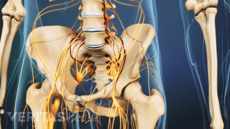
The coccygeal plexus of nerves primarily supplies the pelvis, buttocks, and lower limbs.
The coccyx is innervated by several nerves that transmit sensory information from the pelvic area to the brain.
The coccyx is primarily innervated by the coccygeal plexus, a network of nerves formed by the S5 spinal nerve, the coccygeal ventral rami, and a small descending branch from the S4 spinal nerve.5Apaydin N. Chapter 47 - Variations of the Lumbar and Sacral Plexuses and Their Branches. In: Tubbs RS, Rizk E, Shoja MM, Loukas M, Barbaro N, Spinner RJ, eds. Nerves and Nerve Injuries. Academic Press; 2015. p. 627-645. ISBN 9780124103900. doi:10.1016/B978-0-12-410390-0.00049-4. These nerves primarily supply the pelvis, buttocks, and lower limbs.6Nathan ST, Fisher BE, Roberts CS. Coccydynia. J Bone Joint Surg Br. 2010;92-B(12):1622-1627. doi:10.1302/0301-620X.92B12.25486
Nerves within the coccygeal plexus that innervate the coccyx include:
- Coccygeal nerves. The coccygeal nerves are small nerves that arise from the coccygeal plexus and provide sensory information to the coccyx.7Bordoni B, Sugumar K, Leslie SW. Anatomy, Abdomen and Pelvis, Pelvic Floor. In: StatPearls. Treasure Island (FL): StatPearls Publishing; 2022. PMID: 29489277.
- Dorsal rami. The dorsal rami are small branches of the spinal nerves that emerge from the spinal column and provide sensory information to the skin and muscles of the lower back.8Protzer L, Seligson D, Doursounian L. The coccyx in clinical medicine. Current Orthopaedic Practice. 2017;28(3):314-317. DOI: 10.1097/BCO.0000000000000502
- Somatic nerves. The somatic nerves are responsible for transmitting sensory information from the skin and muscles of the coccyx, helping to detect pain and pressure in the tailbone region.8Protzer L, Seligson D, Doursounian L. The coccyx in clinical medicine. Current Orthopaedic Practice. 2017;28(3):314-317. DOI: 10.1097/BCO.0000000000000502
When the coccyx is injured, it can cause irritation or compression of the nerves, resulting in coccyx pain.1Foye PM. Coccydynia: Tailbone Pain. Phys Med Rehabil Clin N Am. 2017;28(3):539-549. doi:10.1016/j.pmr.2017.03.006 Nerves are delicate, and injuries to them can disrupt signals to and from the brain, leading to improper muscle function and loss of sensation.
Blood Supply to the Coccyx

A network of arteries supplies the coccyx.
The blood supply to the coccyx is primarily derived from the branches of the internal iliac arteries, which are the major arteries that supply the pelvic region.9Mostafa E, Varacallo M. Anatomy, Back, Coccygeal Vertebrae. [Updated 2022 Jun 7]. In: StatPearls [Internet]. Treasure Island (FL): StatPearls Publishing; 2023 Jan-. Available from: https://www.ncbi.nlm.nih.gov/books/NBK549870,10de Assis AM, Moreira AM, de Paula Rodrigues VC, et al. Pelvic Arterial Anatomy Relevant to Prostatic Artery Embolisation and Proposal for Angiographic Classification. Cardiovasc Intervent Radiol. 2015;38(4):855-861. doi:10.1007/s00270-015-1114-3
Other smaller arteries include:
- Middle sacral artery. The middle sacral artery arises from the back of the abdominal aorta and travels down in front of the sacrum to supply the coccyx.7Bordoni B, Sugumar K, Leslie SW. Anatomy, Abdomen and Pelvis, Pelvic Floor. In: StatPearls. Treasure Island (FL): StatPearls Publishing; 2022. PMID: 29489277.
- Lateral sacral arteries. The lateral sacral arteries, which are branches of the internal iliac arteries, send smaller branches to the coccyx and run on the anterior surface of the sacrum and coccyx.
- Superior and inferior gluteal arteries. These arteries are also branches of the internal iliac artery and supply the gluteal region and send branches to the lower part of the sacrum and coccyx.11Lai J, du Plessis M, Wooten C, et al. The blood supply to the sacrotuberous ligament. Surg Radiol Anat. 2017;39(9):953-959. doi:10.1007/s00276-017-1830-2.
Typically, the arteries surrounding the coccyx are protected due to their location being deep in the pelvis; however, they may get damaged from a fall or traumatic injury, leading to tailbone pain (coccydynia) in rare cases.2Mabrouk A, Alloush A, Foye P. Coccyx Pain. [Updated 2023 May 1]. In: StatPearls [Internet]. Treasure Island (FL): StatPearls Publishing; 2023 Jan-. Available from: https://www.ncbi.nlm.nih.gov/books/NBK563139/
Functions of the Coccyx

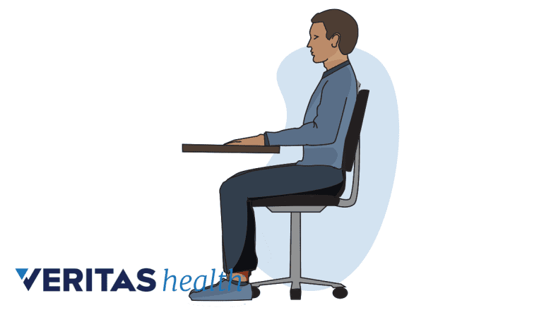
The coccyx helps support the pelvis while sitting.
The coccyx plays a crucial role in the stability of the pelvis and supports several physiological functions.
Major functions of the coccyx include:
- Support and stabilize the body while sitting. The coccyx and the ischial tuberosities work together to support and distribute the body while sitting and leaning back.8Protzer L, Seligson D, Doursounian L. The coccyx in clinical medicine. Current Orthopaedic Practice. 2017;28(3):314-317. DOI: 10.1097/BCO.0000000000000502 The ischial tuberosities are rounded protrusions of bone that make up the bottom of the pelvis. Functionally, a tripod is formed by the two sitting bones and the coccyx (in the midline).2Mabrouk A, Alloush A, Foye P. Coccyx Pain. [Updated 2023 May 1]. In: StatPearls [Internet]. Treasure Island (FL): StatPearls Publishing; 2023 Jan-. Available from: https://www.ncbi.nlm.nih.gov/books/NBK563139/ This tripod supports weight-bearing in the seated position.2Mabrouk A, Alloush A, Foye P. Coccyx Pain. [Updated 2023 May 1]. In: StatPearls [Internet]. Treasure Island (FL): StatPearls Publishing; 2023 Jan-. Available from: https://www.ncbi.nlm.nih.gov/books/NBK563139/
- Serve as an attachment site for the soft tissues of the pelvic floor. The coccyx serves as an anchor point for several muscles and ligaments in the pelvis that make up the pelvic floor, which help support the bladder, rectum, and uterus in females.8Protzer L, Seligson D, Doursounian L. The coccyx in clinical medicine. Current Orthopaedic Practice. 2017;28(3):314-317. DOI: 10.1097/BCO.0000000000000502
- Aid in childbirth. During childbirth, the coccyx can flex or move backward to allow more space for the baby to pass through the birth canal.8Protzer L, Seligson D, Doursounian L. The coccyx in clinical medicine. Current Orthopaedic Practice. 2017;28(3):314-317. DOI: 10.1097/BCO.0000000000000502
The position and alignment of the coccyx can significantly influence the tension and function of the pelvic floor muscles. If the coccyx is misaligned or injured, it can lead to pelvic floor dysfunction that can cause problems with bladder or bowel control, sexual dysfunction, and chronic pain.7Bordoni B, Sugumar K, Leslie SW. Anatomy, Abdomen and Pelvis, Pelvic Floor. In: StatPearls. Treasure Island (FL): StatPearls Publishing; 2022. PMID: 29489277.
Tailbone Pain: Causes, Symptoms, and Risk Factors
Coccydynia, also known as tailbone pain or coccyx pain, is a medical condition characterized by discomfort and pain in the coccyx area.
Coccydynia causes

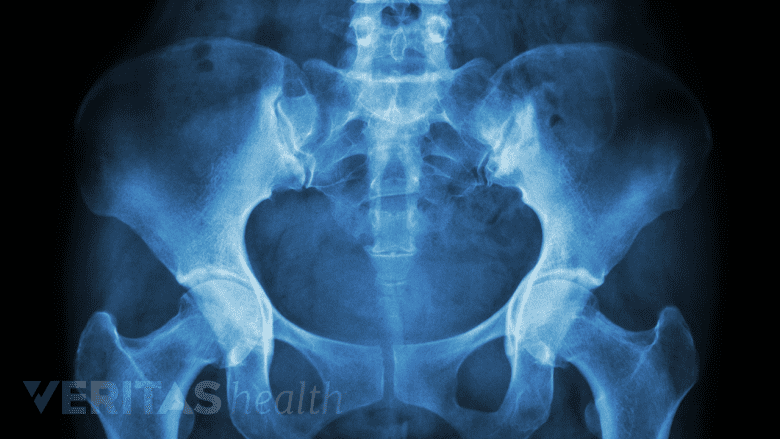
A sudden fall may cause injury to the coccyx.
Traumatic injuries can cause the coccyx to be abnormally displaced forward or backward, become unstable, or get dislocated, leading to painful symptoms.2Mabrouk A, Alloush A, Foye P. Coccyx Pain. [Updated 2023 May 1]. In: StatPearls [Internet]. Treasure Island (FL): StatPearls Publishing; 2023 Jan-. Available from: https://www.ncbi.nlm.nih.gov/books/NBK563139/ The most common causes of such injuries to the coccyx are2Mabrouk A, Alloush A, Foye P. Coccyx Pain. [Updated 2023 May 1]. In: StatPearls [Internet]. Treasure Island (FL): StatPearls Publishing; 2023 Jan-. Available from: https://www.ncbi.nlm.nih.gov/books/NBK563139/:
- Direct vertical trauma (falling on the buttocks)
- Repetitive microtrauma
- Childbirth
Less commonly, infections and tumors may cause coccydynia in some individuals.
In some cases, non-traumatic, repetitive injuries to the coccyx and surrounding soft tissues can occur from sitting for long durations with poor posture, which are telltale signs of fatigue.
Read more about Coccydynia Causes
Coccydynia symptoms

Sitting in a reclining position may aggravate tailbone pain.
The hallmark symptom of coccydynia is severe, debilitating pain and tenderness felt at the top of the tailbone (upper part of the buttock area).2Mabrouk A, Alloush A, Foye P. Coccyx Pain. [Updated 2023 May 1]. In: StatPearls [Internet]. Treasure Island (FL): StatPearls Publishing; 2023 Jan-. Available from: https://www.ncbi.nlm.nih.gov/books/NBK563139/ The pain becomes worse while sitting, especially when sitting in a reclined position. Standing up from a seated position momentarily exacerbates the pain.
Coccydynia risk factors

A broader pelvis in women makes them more prone to coccygeal injuries.
Research indicates that women are five times more likely to develop coccydynia than men.12Lirette LS, Chaiban G, Tolba R, Eissa H. Coccydynia: an overview of the anatomy, etiology, and treatment of coccyx pain. Ochsner J. 2014;14(1):84‐87. Women are typically considered at higher risk for coccydynia because:
- Women have a broader pelvic structure, which may decrease the amount of pelvic rotation, exposing the coccyx to injury.8Protzer L, Seligson D, Doursounian L. The coccyx in clinical medicine. Current Orthopaedic Practice. 2017;28(3):314-317. DOI: 10.1097/BCO.0000000000000502
- Women tend to place more weight on the coccyx when sitting, which makes it more susceptible to injury.1Foye PM. Coccydynia: Tailbone Pain. Phys Med Rehabil Clin N Am. 2017;28(3):539-549. doi:10.1016/j.pmr.2017.03.006
- The natural childbirth process may cause acute damage to the ligaments surrounding the coccyx or the coccyx itself as the baby moves through the birth canal.1Foye PM. Coccydynia: Tailbone Pain. Phys Med Rehabil Clin N Am. 2017;28(3):539-549. doi:10.1016/j.pmr.2017.03.006
Pelvic muscle cramps can also play a role in increased coccyx pain in women. In physical evaluations, women have reported significantly increased coccyx pain during the premenstrual period.1Foye PM. Coccydynia: Tailbone Pain. Phys Med Rehabil Clin N Am. 2017;28(3):539-549. doi:10.1016/j.pmr.2017.03.006
Tailbone pain typically resolves with or without medical treatment. Research indicates that up to 90% of people find relief from coccydynia symptoms with nonsurgical care.2Mabrouk A, Alloush A, Foye P. Coccyx Pain. [Updated 2023 May 1]. In: StatPearls [Internet]. Treasure Island (FL): StatPearls Publishing; 2023 Jan-. Available from: https://www.ncbi.nlm.nih.gov/books/NBK563139/ In rare cases, if medical treatment is delayed, the condition may become chronic and cause severe pain and functional impairment in some individuals.2Mabrouk A, Alloush A, Foye P. Coccyx Pain. [Updated 2023 May 1]. In: StatPearls [Internet]. Treasure Island (FL): StatPearls Publishing; 2023 Jan-. Available from: https://www.ncbi.nlm.nih.gov/books/NBK563139/
Read about Treatment for Coccydynia (Tailbone Pain)
- 1 Foye PM. Coccydynia: Tailbone Pain. Phys Med Rehabil Clin N Am. 2017;28(3):539-549. doi:10.1016/j.pmr.2017.03.006
- 2 Mabrouk A, Alloush A, Foye P. Coccyx Pain. [Updated 2023 May 1]. In: StatPearls [Internet]. Treasure Island (FL): StatPearls Publishing; 2023 Jan-. Available from: https://www.ncbi.nlm.nih.gov/books/NBK563139/
- 3 Sandrasegaram N, Gupta R, Baloch M. Diagnosis and management of sacrococcygeal pain. BJA Educ. 2020;20(3):74-79. doi:10.1016/j.bjae.2019.11.004
- 4 Hauser RA, Dolan EE, Phillips HJ, Newlin AC, Morre RE, Woldin BA. Ligament Injury and Healing: A Review of Current Clinical Diagnostics and Therapeutics. The Open Rehabilitation Journal. 2013;6:1-20. doi:10.2174/1874943701306010001
- 5 Apaydin N. Chapter 47 - Variations of the Lumbar and Sacral Plexuses and Their Branches. In: Tubbs RS, Rizk E, Shoja MM, Loukas M, Barbaro N, Spinner RJ, eds. Nerves and Nerve Injuries. Academic Press; 2015. p. 627-645. ISBN 9780124103900. doi:10.1016/B978-0-12-410390-0.00049-4.
- 6 Nathan ST, Fisher BE, Roberts CS. Coccydynia. J Bone Joint Surg Br. 2010;92-B(12):1622-1627. doi:10.1302/0301-620X.92B12.25486
- 7 Bordoni B, Sugumar K, Leslie SW. Anatomy, Abdomen and Pelvis, Pelvic Floor. In: StatPearls. Treasure Island (FL): StatPearls Publishing; 2022. PMID: 29489277.
- 8 Protzer L, Seligson D, Doursounian L. The coccyx in clinical medicine. Current Orthopaedic Practice. 2017;28(3):314-317. DOI: 10.1097/BCO.0000000000000502
- 9 Mostafa E, Varacallo M. Anatomy, Back, Coccygeal Vertebrae. [Updated 2022 Jun 7]. In: StatPearls [Internet]. Treasure Island (FL): StatPearls Publishing; 2023 Jan-. Available from: https://www.ncbi.nlm.nih.gov/books/NBK549870
- 10 de Assis AM, Moreira AM, de Paula Rodrigues VC, et al. Pelvic Arterial Anatomy Relevant to Prostatic Artery Embolisation and Proposal for Angiographic Classification. Cardiovasc Intervent Radiol. 2015;38(4):855-861. doi:10.1007/s00270-015-1114-3
- 11 Lai J, du Plessis M, Wooten C, et al. The blood supply to the sacrotuberous ligament. Surg Radiol Anat. 2017;39(9):953-959. doi:10.1007/s00276-017-1830-2.
- 12 Lirette LS, Chaiban G, Tolba R, Eissa H. Coccydynia: an overview of the anatomy, etiology, and treatment of coccyx pain. Ochsner J. 2014;14(1):84‐87.

