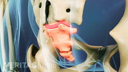
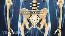
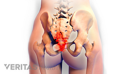
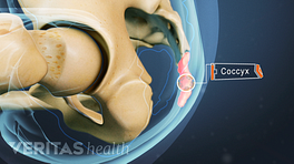
The coccyx is a triangular arrangement of bone that makes up the very bottom portion of the spine below the sacrum. It is the final segment of the vertebral column and represents a vestigial tail, hence the common term tailbone.
The coccyx is an integral part of the pelvis, which is vital to essential activities like moving from sitting to standing, walking, eliminating waste, sexual intercourse, and childbirth.
The word coccyx is derived from Greek and means “beak,” as it resembles the shape of a cuckoo bird’s beak when viewed from the side.
In This Article:
- Anatomy of the Coccyx (Tailbone)
- Soft Tissues and Essential Functions of the Coccyx
- Coccydynia (Tailbone Pain) Video
Understanding the Coccyx: Location, Structure, and Fusion
The coccyx is located at the very end of the spine and consists of approximately 4 fused vertebrae.
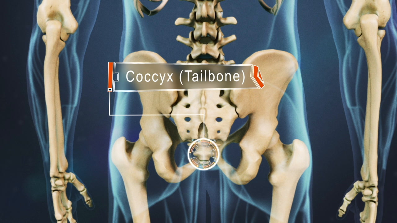
The coccyx comprises 3-5 fused coccygeal vertebrae that get progressively smaller from top to bottom. The entire coccyx measures approximately 4-10 cm in length and is positioned as an inverted triangle—with the base on top and the apex below. 1 Protzer L, Seligson D, Doursounian L. The coccyx in clinical medicine. Current Orthopaedic Practice. 2017;28(3):314-317. DOI: 10.1097/BCO.0000000000000502
When viewed from the side, the coccyx has varying degrees of a convex angle, appearing in the direction of the thoracic spine or middle back.
The parts of the coccyx are discussed below 2 Foye PM. Coccydynia: Tailbone Pain. Phys Med Rehabil Clin N Am. 2017;28(3):539-549. doi:10.1016/j.pmr.2017.03.006 :
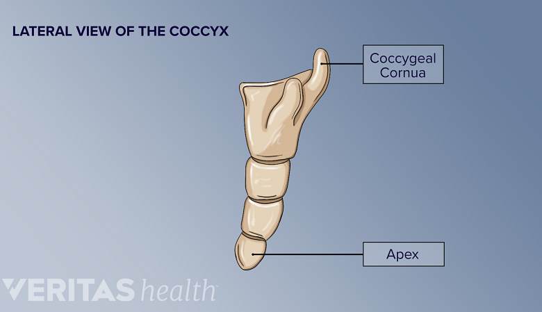
- Base. The base of the coccyx is formed by the first and broadest coccygeal vertebra, Co1, which sits immediately below S5, the 5th sacral vertebra. Co1 is the only coccygeal vertebra with the following two unique features:
- Transverse processes: bony attachment points for pelvic soft tissues
- Coccygeal cornua: two horn-like structures that project upward from the base of the coccyx. The coccygeal cornua articulate with the cornua of the sacrum, forming part of the sacrococcygeal symphysis joint and the intervertebral foramen that the S5 nerve passes through.
- Posterior surface. The coccyx's posterior (back) surface contains small, rounded protrusions that are remnants of the spinous processes of the fused coccygeal vertebrae.
- Anterior surface. The coccyx's anterior (front) surface contains slightly concave grooves that are remnants of the vertebral foramina.
- Lateral surface. The lateral surfaces or sides of the coccyx serve as points of attachment for nearby soft tissues.
- Apex. The apex of the coccyx is the pointed end, which curves forward and downward. The apex is a point of attachment for soft tissues that support the pelvic floor and anus.
Despite its small size, the coccyx is crucial in providing stability for the lower back and pelvis,1 the weight-bearing tripod structure that supports a sitting position.
The coccygeal vertebrae fuse in early adulthood
During early development, each vertebra of the coccyx forms independently, similar to the rest of the vertebral column.
Through adolescence and early adulthood, the vertebrae gradually fuse to form a solid bone structure through a process known as coccygeal ossification. 3 Woon JTK, Stringer MD. Clinical anatomy of the coccyx: A systematic review. Clinical Anatomy. 2012;25(12):158-167. https://doi.org/10.1002/ca.21216 The timing of coccygeal ossification varies among individuals, but it is typically complete by the mid-20s. 3 Woon JTK, Stringer MD. Clinical anatomy of the coccyx: A systematic review. Clinical Anatomy. 2012;25(12):158-167. https://doi.org/10.1002/ca.21216
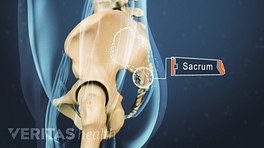
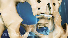
Sacrococcygeal Symphysis: The Articulation between the Sacrum and the Coccyx
The sacrococcygeal symphysis is a small intervertebral joint that connects S5 to Co1. 4 Chaudhry SR, Nahian A, Chaudhry K. Anatomy, Abdomen and Pelvis, Pelvis. In: StatPearls. StatPearls Publishing, Treasure Island (FL); 2022. https://europepmc.org/article/NBK/nbk482258
Structure of the sacrococcygeal symphysis
The sacrococcygeal symphysis is a slightly moveable joint. The joint contributes to pelvic stability and is strengthened by the tough and resilient set of anterior, posterior, and lateral sacrococcygeal ligaments. 1 Protzer L, Seligson D, Doursounian L. The coccyx in clinical medicine. Current Orthopaedic Practice. 2017;28(3):314-317. DOI: 10.1097/BCO.0000000000000502
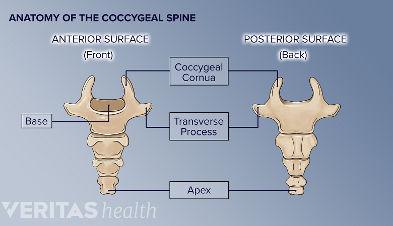
Sacrococcygeal disc
The sacrococcygeal disc serves as a cushion to absorb weight from the upper body.
A thin fibro-cartilaginous disc is situated between S5 and Co1, and it serves as a cushion to absorb weight from the upper body. 4 Chaudhry SR, Nahian A, Chaudhry K. Anatomy, Abdomen and Pelvis, Pelvis. In: StatPearls. StatPearls Publishing, Treasure Island (FL); 2022. https://europepmc.org/article/NBK/nbk482258 This disc is thinner and less robust than the other vertebral discs in the spinal column. 4 Chaudhry SR, Nahian A, Chaudhry K. Anatomy, Abdomen and Pelvis, Pelvis. In: StatPearls. StatPearls Publishing, Treasure Island (FL); 2022. https://europepmc.org/article/NBK/nbk482258
Like other spinal discs, the sacrococcygeal disc may degenerate due to natural aging. However, it usually does not encounter herniation as it is exposed to lower mechanical stress levels than the other spinal discs. 5 Sandrasegaram N, Gupta R, Baloch M. Diagnosis and management of sacrococcygeal pain. BJA Educ. 2020;20(3):74-79. doi:10.1016/j.bjae.2019.11.004
Motion at the sacrococcygeal symphysis
The movement of the sacrococcygeal symphysis is limited to bending forward and backward. The joint helps in spinal flexion and extension, such as moving from standing to sitting. 5 Sandrasegaram N, Gupta R, Baloch M. Diagnosis and management of sacrococcygeal pain. BJA Educ. 2020;20(3):74-79. doi:10.1016/j.bjae.2019.11.004
While sitting, the coccyx moves slightly forward, allowing the body’s weight to be distributed evenly. 4 Chaudhry SR, Nahian A, Chaudhry K. Anatomy, Abdomen and Pelvis, Pelvis. In: StatPearls. StatPearls Publishing, Treasure Island (FL); 2022. https://europepmc.org/article/NBK/nbk482258
During childbirth, the coccyx moves backward, increasing the diameter of the pelvic passage to aid in the delivery of a child. 4 Chaudhry SR, Nahian A, Chaudhry K. Anatomy, Abdomen and Pelvis, Pelvis. In: StatPearls. StatPearls Publishing, Treasure Island (FL); 2022. https://europepmc.org/article/NBK/nbk482258 In some cases, the mother’s coccyx may fracture during labor and delivery.
Key Differences Between Coccygeal Vertebrae and Other Spinal Vertebrae
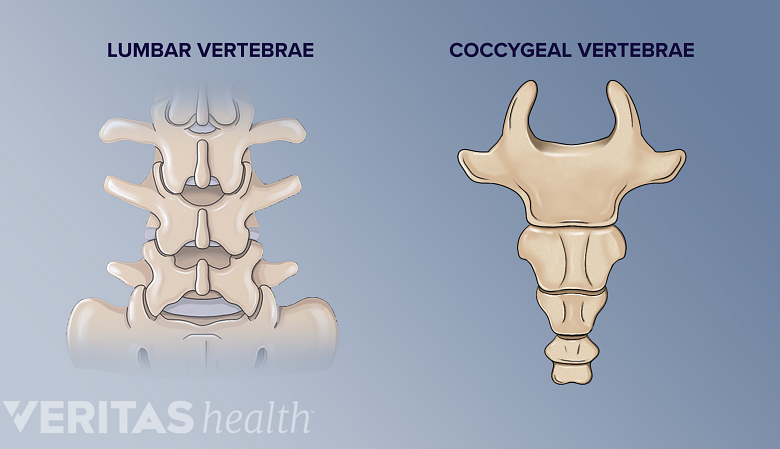
Coccygeal and spinal vertebrae share some similarities but differ significantly.
The coccygeal vertebrae and spinal vertebrae have some similarities, but they also have significant differences.
- Each bone in the coccyx is somewhat smaller in both height and width compared to the bone above it.
- The coccyx does not have an opening for the vertebral column; hence, there are no neural elements, such as the cauda equina or cerebrospinal fluid, in the coccygeal spine. 6 Mostafa E, Varacallo M. Anatomy, Back, Coccygeal Vertebrae. [Updated 2022 Jun 7]. In: StatPearls [Internet]. Treasure Island (FL): StatPearls Publishing; 2023 Jan-. Available from: https://www.ncbi.nlm.nih.gov/books/NBK549870/
Only the first and largest coccygeal vertebra, Co1, resemble other spinal vertebrae because of its transverse and superior articular processes. 6 Mostafa E, Varacallo M. Anatomy, Back, Coccygeal Vertebrae. [Updated 2022 Jun 7]. In: StatPearls [Internet]. Treasure Island (FL): StatPearls Publishing; 2023 Jan-. Available from: https://www.ncbi.nlm.nih.gov/books/NBK549870/
Anatomical Variations of the Coccyx
The coccyx generally follows a slight forward curve.
Anatomical variations of the coccyx are common.Common variations reported in the literature include:
- Differences in the number of segments. According to the literature, around 76% of individuals have four coccygeal vertebrae, but some can have as few as three (13%) or as many as five (11%).
7
Woon JT, Perumal V, Maigne JY, Stringer MD. CT morphology and morphometry of the normal adult coccyx. Eur Spine J. 2013;22(4):863-870. doi:10.1007/s00586-012-2595-2
Rarely, some individuals may have only two fused segments (hypoplastic coccyx), while others may have more than five segments due to an extra coccygeal vertebra (supernumerary vertebra). 8 Paudyal S, Karna BP, Joshi BR. Evaluation of Anatomical Variation of Human Coccyx on Magnetic Resonance Imaging. Nepalese J Radiol. 2022;12(2):2-6. doi:10.3126/njr.v12i2.54582. - Changes in the degree of curvature. The curvature of the coccyx generally follows a slight forward curve, but some individuals may have a more exaggerated forward curve or have a straighter or even a slightly backward-curving coccyx. 3 Woon JTK, Stringer MD. Clinical anatomy of the coccyx: A systematic review. Clinical Anatomy. 2012;25(12):158-167. https://doi.org/10.1002/ca.21216 The average coccygeal curve length in males and females combined is 4.3 cm, and the mean coccygeal angle is 143°. 3 Woon JTK, Stringer MD. Clinical anatomy of the coccyx: A systematic review. Clinical Anatomy. 2012;25(12):158-167. https://doi.org/10.1002/ca.21216
- The presence of bony projections. While less common, some individuals may have bony projections or spurs on the coccyx. 2 Foye PM. Coccydynia: Tailbone Pain. Phys Med Rehabil Clin N Am. 2017;28(3):539-549. doi:10.1016/j.pmr.2017.03.006
- Atypical fusion of the coccygeal vertebrae. Some people may have only partial fusions between the coccygeal segments. 8 Paudyal S, Karna BP, Joshi BR. Evaluation of Anatomical Variation of Human Coccyx on Magnetic Resonance Imaging. Nepalese J Radiol. 2022;12(2):2-6. doi:10.3126/njr.v12i2.54582.
- The absence of the coccyx. In rare cases, some individuals may not develop the coccyx. This condition is called coccygeal agenesis. 8 Paudyal S, Karna BP, Joshi BR. Evaluation of Anatomical Variation of Human Coccyx on Magnetic Resonance Imaging. Nepalese J Radiol. 2022;12(2):2-6. doi:10.3126/njr.v12i2.54582. , 9 Lee JY, Pang D, Wang KC. Caudal Agenesis and Associated Spinal Cord Malformations. In: Di Rocco C, Pang D, Rutka J, eds. Textbook of Pediatric Neurosurgery. Springer; 2020. https://doi.org/10.1007/978-3-319-72168-2_119
It is important to note that while anatomical variations in the coccyx are relatively common, they typically do not cause significant functional issues. However, in some cases, certain variations or abnormalities like coccygeal agenesis or bony projection may contribute to discomfort, pain, or other coccyx-related conditions. 2 Foye PM. Coccydynia: Tailbone Pain. Phys Med Rehabil Clin N Am. 2017;28(3):539-549. doi:10.1016/j.pmr.2017.03.006 , 9 Lee JY, Pang D, Wang KC. Caudal Agenesis and Associated Spinal Cord Malformations. In: Di Rocco C, Pang D, Rutka J, eds. Textbook of Pediatric Neurosurgery. Springer; 2020. https://doi.org/10.1007/978-3-319-72168-2_119
Read more about Coccydynia (Tailbone Pain)
- 1 Protzer L, Seligson D, Doursounian L. The coccyx in clinical medicine. Current Orthopaedic Practice. 2017;28(3):314-317. DOI: 10.1097/BCO.0000000000000502
- 2 Foye PM. Coccydynia: Tailbone Pain. Phys Med Rehabil Clin N Am. 2017;28(3):539-549. doi:10.1016/j.pmr.2017.03.006
- 3 Woon JTK, Stringer MD. Clinical anatomy of the coccyx: A systematic review. Clinical Anatomy. 2012;25(12):158-167. https://doi.org/10.1002/ca.21216
- 4 Chaudhry SR, Nahian A, Chaudhry K. Anatomy, Abdomen and Pelvis, Pelvis. In: StatPearls. StatPearls Publishing, Treasure Island (FL); 2022. https://europepmc.org/article/NBK/nbk482258
- 5 Sandrasegaram N, Gupta R, Baloch M. Diagnosis and management of sacrococcygeal pain. BJA Educ. 2020;20(3):74-79. doi:10.1016/j.bjae.2019.11.004
- 6 Mostafa E, Varacallo M. Anatomy, Back, Coccygeal Vertebrae. [Updated 2022 Jun 7]. In: StatPearls [Internet]. Treasure Island (FL): StatPearls Publishing; 2023 Jan-. Available from: https://www.ncbi.nlm.nih.gov/books/NBK549870/
- 7 Woon JT, Perumal V, Maigne JY, Stringer MD. CT morphology and morphometry of the normal adult coccyx. Eur Spine J. 2013;22(4):863-870. doi:10.1007/s00586-012-2595-2
- 8 Paudyal S, Karna BP, Joshi BR. Evaluation of Anatomical Variation of Human Coccyx on Magnetic Resonance Imaging. Nepalese J Radiol. 2022;12(2):2-6. doi:10.3126/njr.v12i2.54582.
- 9 Lee JY, Pang D, Wang KC. Caudal Agenesis and Associated Spinal Cord Malformations. In: Di Rocco C, Pang D, Rutka J, eds. Textbook of Pediatric Neurosurgery. Springer; 2020. https://doi.org/10.1007/978-3-319-72168-2_119