
Subscribe to our newsletter and get your FREE guide,
Natural Back Pain Relief: 16 Choices for Lasting Comfort.
Collectively, the vertebral bodies make up the bony building blocks of the spine. The bones are stacked on top of each other and securely attached with a disc in between each one.

The spinal column consists of cervical, thoracic, and lumbar vertebrae.
The vertebral bodies act as a support column to hold up the spine. This column supports about half of the weight of the body, with the other half supported by the muscles.
Each area of the spine has some differences in the form and function of vertebral bodies and how they are attached to adjacent structures, as follows:
In This Article:
- Spinal Anatomy and Back Pain
- Vertebrae in the Vertebral Column
- Spinal Discs
- Spinal Cord and Spinal Nerve Roots
- Back Muscles and Low Back Pain
- Sacrum (Sacral Region)
- Spine Anatomy Overview Video
The cervical spine (neck) has seven vertebrae. The first two vertebrae are unique:
- The first cervical vertebra, called the atlas, is shaped like a ring. This vertebra is named C1.
- The second vertebral body is called the axis and acts like a post that supports the atlas.
- Like the rest of the spine, the next five vertebrae (C3-C7) have three joints at each segment, including one disc in the front and a pair of facet joints in the back.
advertisement
Unlike the rest of the spine, the segments in the cervical spine contain openings in each vertebral body for arteries to carry blood to the brain (for exampe, vertebral artery runs through the transverse foramen).
The cervical vertebrae also serve an important function in protecting the spinal cord, which travels through a space in the center of the vertebral bones and connects the brain with the rest of the body.
Vertebrae in the Thoracic Spine

The thoracic spine has 12 vertebrae.
The thoracic spine makes up the upper back and has twelve vertebral bodies, labeled T1 through T12.
These structures have very little motion because they are firmly attached to the ribs and sternum (breastbone). There is little motion in the thoracic spine, so this region has less risk of injury or wear and tear.
Since they are major load bearing structures, vertebral bodies in the upper back are prone to developing compression fractures, particularly in patients with osteoporosis (a condition that weakens the bone). These fractures can lead to chronic back pain and progressive misalignment or deformity of the spine.
See When Back Pain Is a Spine Compression Fracture
Other areas of the upper back can also be a source of pain, it is just less common than in the neck or lower back.
advertisement
Vertebrae of the Lumbar Spine
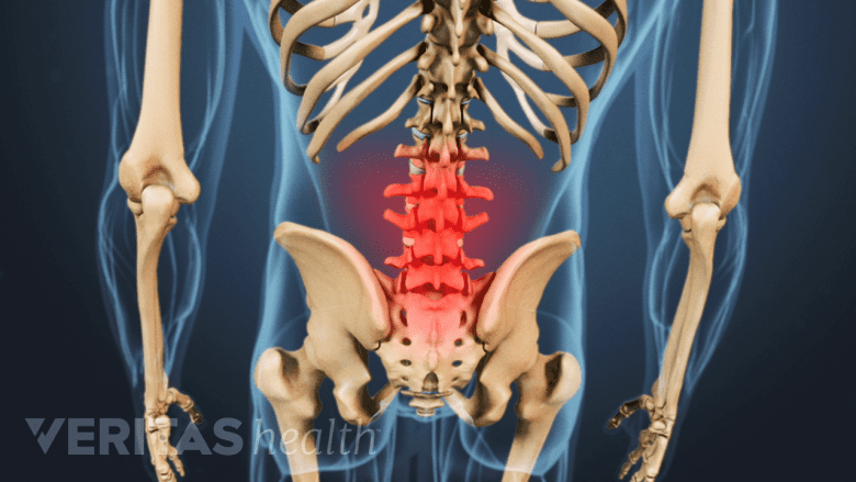
The lumbar spine has 5 large vertebrae.
The lumbar spine has five vertebrae, labeled L1-L5, that extend from the lower thoracic spine to the sacrum at bottom of the spine. The vertebrae of the lower back are the largest of the spine and they bear the majority of the body's weight.
Approximately 50% of flexion (bending forward) occurs at the hips, and 50% occurs at the lumbar spine (lower back).
Each segment of the lumbar spine comprises the following structures:
- The thick oval segment of bone in the front of the spinal column is called the vertebral body.
- The vertebral bodies are attached to a bony arch through which all the nerve roots pass as they exit the spinal column.
- The vertebral arches are interconnected by paired facet joints, which in combination with the disc, create a three-joint complex at each vertebral motion segment. This complex provides spinal support and allows for motion.
- The facet joints have cartilage on the joint surfaces and a capsule around them. The cartilage can degenerate as one ages, and lead to degenerative arthritis.
- The spinous process protrudes from the back of the spine, and these are the bony ridges that can be felt through the skin along the back of the spine.
Each vertebral segment is made up of two bony vertebrae with a spinal disc in between. The spinal disc is also susceptible to a number of problems and may often be a source of pain, as discussed on the next page.

advertisement