
Subscribe to our newsletter and get your FREE guide,
Natural Back Pain Relief: 16 Choices for Lasting Comfort.
The sciatic nerve is the largest and longest nerve in the human body, originating at the base of the spine and running along the back of each leg into the foot.1Davis D, Vasudevan A. Sciatica. [Updated 2019 Feb 28]. In: StatPearls [Internet]. Treasure Island (FL): StatPearls Publishing; 2019 Jan-. Available from: https://www.ncbi.nlm.nih.gov/books/NBK507908/,2Davis D, Vasudevan A. Sciatica. [Updated 2019 Feb 28]. In: StatPearls [Internet]. Treasure Island (FL): StatPearls Publishing; 2019 Jan-. Available from: https://www.ncbi.nlm.nih.gov/books/NBK507908/ At its thickest point, it is about as wide as an adult thumb.
In This Article:
- Sciatic Nerve Anatomy
- Sciatic Nerve: Muscle Innervation and Function
- Sciatic Nerve Anatomy Video
A Comprehensive Overview of the Sciatic Nerve
The sciatic nerve is formed in the lower spine by the combination of motor and sensory fibers from spinal nerves L4 to S3. These spinal nerves belong to a larger group of nerves in the lower spine called the lumbosacral plexus.
This long, thick, and bulky nerve runs along the back of the thigh and leg and terminates in the foot.
The nerve supplies most areas of the thigh, leg, and foot.3Barral J, Croibier A. Manual Therapy for the Peripheral Nerves. Elsevier Health Sciences; 2007.
advertisement
The mixed (sensory and motor) sciatic nerve provides the majority of the functions in the lower limbs and makes actions such as walking, running, climbing, lifting weights, and standing possible.
Location of the Sciatic Nerve in the Body
The sciatic nerve extends from the lower back to the foot and is a mixed nerve, which means it has both motor and sensory fibers. These fibers provide sensation and function in the lower limbs.
The Origin and Course of the Sciatic Nerve: Tracing its Pathway

The sciatic nerve is formed in the lower spine by the combination of spinal nerves L4 to S3.
A combination of 5 nerve roots that exit from inside the lower lumbar and upper sacral spine—L4, L5, S1, S2, and S3—forms the sciatic nerve. These 5 nerves group together deep in the buttock, near the front surface of the piriformis muscle, and combine to form the single large, thick sciatic nerve.1Davis D, Vasudevan A. Sciatica. [Updated 2019 Feb 28]. In: StatPearls [Internet]. Treasure Island (FL): StatPearls Publishing; 2019 Jan-. Available from: https://www.ncbi.nlm.nih.gov/books/NBK507908/,4Ryan MM, Jones HR Jr. Mononeuropathies. In: Neuromuscular Disorders of Infancy, Childhood, and Adolescence. Elsevier; 2015:243-273. doi:10.1016/b978-0-12-417044-5.00014-7
The sciatic nerve is large and round
At its origin in the buttock, the sciatic nerve is shaped like a flattened band that is about 5 mm high and 10 mm to 15 mm wide. As it continues down into the leg, the nerve forms into a more rounded shape.3Barral J, Croibier A. Manual Therapy for the Peripheral Nerves. Elsevier Health Sciences; 2007. At its thickest portion, the nerve measures about 2 cm in diameter,1Davis D, Vasudevan A. Sciatica. [Updated 2019 Feb 28]. In: StatPearls [Internet]. Treasure Island (FL): StatPearls Publishing; 2019 Jan-. Available from: https://www.ncbi.nlm.nih.gov/books/NBK507908/ about the same circumference of a US penny.
The sciatic nerve is the longest nerve in the body
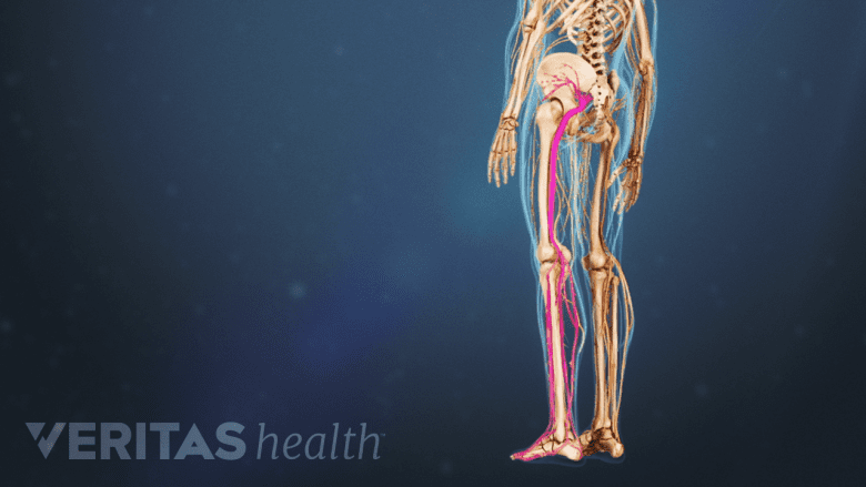
The sciatic nerve is the longest nerve in the body.
In the pelvis, the sciatic nerve and a few other surrounding nerves and blood vessels exit through an opening called the greater sciatic foramen (sciatic notch). This opening is located deep in the buttock, just below the piriformis muscle.4Ryan MM, Jones HR Jr. Mononeuropathies. In: Neuromuscular Disorders of Infancy, Childhood, and Adolescence. Elsevier; 2015:243-273. doi:10.1016/b978-0-12-417044-5.00014-7
- After exiting the greater sciatic foramen, the sciatic nerve rests on the back portion of the ischium—the curved bone at the base of the pelvis.3Barral J, Croibier A. Manual Therapy for the Peripheral Nerves. Elsevier Health Sciences; 2007.
- It then courses down and runs below and along the side of the large gluteus maximus muscle in the buttock.3Barral J, Croibier A. Manual Therapy for the Peripheral Nerves. Elsevier Health Sciences; 2007.
- The nerve descends by crossing behind a combination of muscles located deep in the hip joint.3Barral J, Croibier A. Manual Therapy for the Peripheral Nerves. Elsevier Health Sciences; 2007.
- At the lower edge of the gluteus maximus muscle of the buttock, the nerve reaches the back portion of the upper thigh.3Barral J, Croibier A. Manual Therapy for the Peripheral Nerves. Elsevier Health Sciences; 2007.
- The nerve lies deep within the thigh, covered by the large thigh muscle, called the biceps femoris.3Barral J, Croibier A. Manual Therapy for the Peripheral Nerves. Elsevier Health Sciences; 2007.
The sciatic nerve then progresses downward between the interconnected muscles of the thigh. It is surrounded by a single long fatty sheath from the pelvis to the knee. At the knee, the nerve divides into two branches.
Sciatic Nerve Branches: A Detailed Analysis of its Divisions
The sciatic nerve splits into 2 main branches near the back of the knee at a point called the popliteal fossa. The popliteal fossa is a rhomboid-shaped space that serves as a conduit for blood vessels and nerves in the leg. This fossa is located slightly above the joint fold at the back of the knee.3Barral J, Croibier A. Manual Therapy for the Peripheral Nerves. Elsevier Health Sciences; 2007.
Main branches
At the popliteal fossa:
- The tibial nerve continues down the back of the calf to the heel and sole of the foot.
- The common peroneal nerve (common fibular nerve) travels sideways along the outer part of the knee to the outer border of the lower leg and foot.
Both these nerves finally terminate into small sensory nerves in the calf that innervate the outer side of each foot. These sensory nerves are called the sural nerves.
Smaller branches

The sciatic nerve gives off smaller branches, called collaterals.
Along its course, the sciatic nerve gives off smaller branches, called collaterals, which include the:
- Muscle branches of the sciatic nerve that supply the muscles in the thigh–including the hamstring group at the back of the thigh, and the adductor magnus muscles along the inner thigh.3Barral J, Croibier A. Manual Therapy for the Peripheral Nerves. Elsevier Health Sciences; 2007. Other small branches supply the leg and foot muscles.3Barral J, Croibier A. Manual Therapy for the Peripheral Nerves. Elsevier Health Sciences; 2007.
- Articular branches of the sciatic nerve that supply the back of the hip joint and the back and side of the knee joint.3Barral J, Croibier A. Manual Therapy for the Peripheral Nerves. Elsevier Health Sciences; 2007.
While the sciatic nerve does not supply any structure in the buttock, pain may be referred to this area when the sciatic nerve is impaired.3Barral J, Croibier A. Manual Therapy for the Peripheral Nerves. Elsevier Health Sciences; 2007.
Blood Supply of the Sciatic Nerve

A vast network of blood vessels supplies the sciatic nerve.
The delivery of nutrients to the sciatic nerve is facilitated through an extensive system of blood vessels, which contribute to the nerve’s function. An interruption of blood flow to this nerve can cause pain and dysfunction.
The sciatic nerve and its branches receive their blood supply from the following two sources2Davis D, Vasudevan A. Sciatica. [Updated 2019 Feb 28]. In: StatPearls [Internet]. Treasure Island (FL): StatPearls Publishing; 2019 Jan-. Available from: https://www.ncbi.nlm.nih.gov/books/NBK507908/:
- The extrinsic system, which consists of contributions from nearby arteries and veins.
- The intrinsic system, which includes arteries and veins that run along the nerve and are embedded deep in a sheath of connective tissue that envelops the nerve (epineurium).
The extrinsic and intrinsic systems connect at various junction points. The blood flow within the nerve’s blood vessel system is highly variable and consists of many smaller networks.2Davis D, Vasudevan A. Sciatica. [Updated 2019 Feb 28]. In: StatPearls [Internet]. Treasure Island (FL): StatPearls Publishing; 2019 Jan-. Available from: https://www.ncbi.nlm.nih.gov/books/NBK507908/ The intrinsic blood supply may be affected by conditions such as diabetes, contributing to symptoms associated with diabetic neuropathy.
advertisement
Variations in the Anatomy of the Sciatic Nerve

Different sciatic nerve pathways in the pelvis.
It is estimated that around 16% of the population may have variations in the anatomical structure of the sciatic nerve.2Davis D, Vasudevan A. Sciatica. [Updated 2019 Feb 28]. In: StatPearls [Internet]. Treasure Island (FL): StatPearls Publishing; 2019 Jan-. Available from: https://www.ncbi.nlm.nih.gov/books/NBK507908/,5Ryan MM, Jones HR Jr. Mononeuropathies. In: Neuromuscular Disorders of Infancy, Childhood, and Adolescence. Elsevier; 2015:243-273. doi:10.1016/b978-0-12-417044-5.00014-7 While the variants are considered normal, they may increase the risk of developing sciatica pain due to impingement, entrapment, or irritation of the nerve root.5Ryan MM, Jones HR Jr. Mononeuropathies. In: Neuromuscular Disorders of Infancy, Childhood, and Adolescence. Elsevier; 2015:243-273. doi:10.1016/b978-0-12-417044-5.00014-7
Typical variations in sciatic nerve anatomy are described below2Davis D, Vasudevan A. Sciatica. [Updated 2019 Feb 28]. In: StatPearls [Internet]. Treasure Island (FL): StatPearls Publishing; 2019 Jan-. Available from: https://www.ncbi.nlm.nih.gov/books/NBK507908/:
- The sciatic nerve divides above the piriformis muscle; one portion passes through the piriformis, the other leaves the pelvis below the muscle. This variant is most common.
- The sciatic nerve divides above the piriformis muscle; one portion passes through the piriformis, the other leaves the pelvic area above the muscle.
- The sciatic nerve divides above the piriformis, one portion travels in front of it, the other travels behind it.
- An undivided sciatic nerve exits through the piriformis muscle.
- An undivided sciatic nerve exits from behind the top part of the piriformis.
In the above cases where the sciatic nerve divides, both portions of the nerve immediately merge again and course downward as a single nerve.
In around 10% of the population, the nerve may divide at a level above the popliteal fossa but then does not merge and courses down in two separate branches (some researchers may not consider this possibility as a variant).4Ryan MM, Jones HR Jr. Mononeuropathies. In: Neuromuscular Disorders of Infancy, Childhood, and Adolescence. Elsevier; 2015:243-273. doi:10.1016/b978-0-12-417044-5.00014-7
Sciatic Nerve Pain
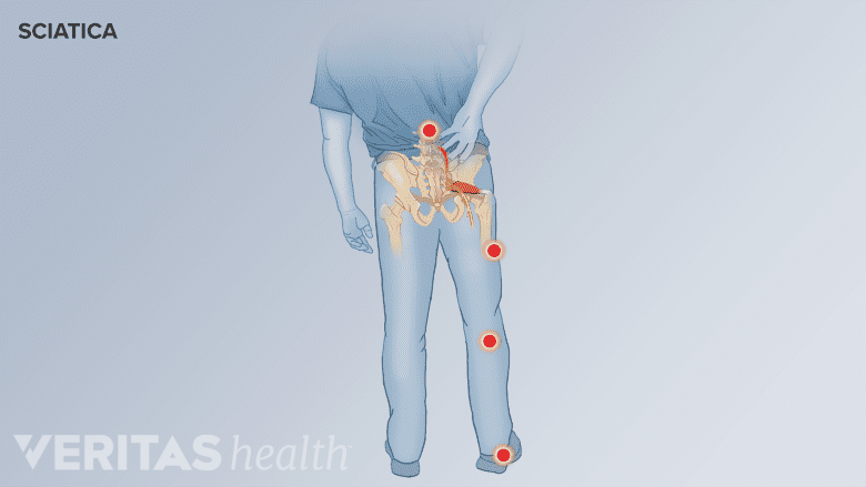
Distribution of sciatic nerve pain.
A healthy sciatic nerve is well protected near its origin by the buttock muscles and cannot be felt by touching or pressing on the skin. When the sciatic nerve is impaired, the leg may feel stiff and inflexible during movements.3Barral J, Croibier A. Manual Therapy for the Peripheral Nerves. Elsevier Health Sciences; 2007. When inflamed or impinged, the nerve can lead to debilitating pain, weakness, and tingling in the lower back, buttock, and leg(s).
Watch Sciatica Causes and Symptoms Video
A problem with the sciatic nerve may occur in the lumbar spine (lower back) at the nerve root level, where the nerve originates. Problems can also occur along the course of the nerve in the thigh, leg, or foot. The symptoms are usually felt in the areas around and below the location where the nerve is affected.
- When the nerve root(s) is affected, the condition is called lumbar radiculopathy and is commonly referred to as sciatica.
- When the body of the nerve is affected along its course, the condition is called sciatic neuropathy.
The symptoms and signs of a sciatic nerve problem are commonly experienced as pain, numbness, altered sensation, and/or weakness that affect the part of the leg supplied by the nerve (right or left). In rare cases, both legs may be affected.
The sciatic nerve is a major component of the human body, supplying motor function to move each leg and foot in multiple directions as well as sensory functions along the path of the nerve and its extensions.
- 1 Davis D, Vasudevan A. Sciatica. [Updated 2019 Feb 28]. In: StatPearls [Internet]. Treasure Island (FL): StatPearls Publishing; 2019 Jan-. Available from: https://www.ncbi.nlm.nih.gov/books/NBK507908/
- 2 Davis D, Vasudevan A. Sciatica. [Updated 2019 Feb 28]. In: StatPearls [Internet]. Treasure Island (FL): StatPearls Publishing; 2019 Jan-. Available from: https://www.ncbi.nlm.nih.gov/books/NBK507908/
- 3 Barral J, Croibier A. Manual Therapy for the Peripheral Nerves. Elsevier Health Sciences; 2007.
- 4 Ryan MM, Jones HR Jr. Mononeuropathies. In: Neuromuscular Disorders of Infancy, Childhood, and Adolescence. Elsevier; 2015:243-273. doi:10.1016/b978-0-12-417044-5.00014-7

advertisement