Your lower back is a superb feat of engineering—it’s strong, weight-bearing, and sturdy, yet highly flexible with a range of motion in all directions.
Understanding the anatomy of your lower spine can help you communicate more effectively with the medical professionals who treat your lower back pain.
Here is a description of useful anatomical landmarks.
The lordotic curve
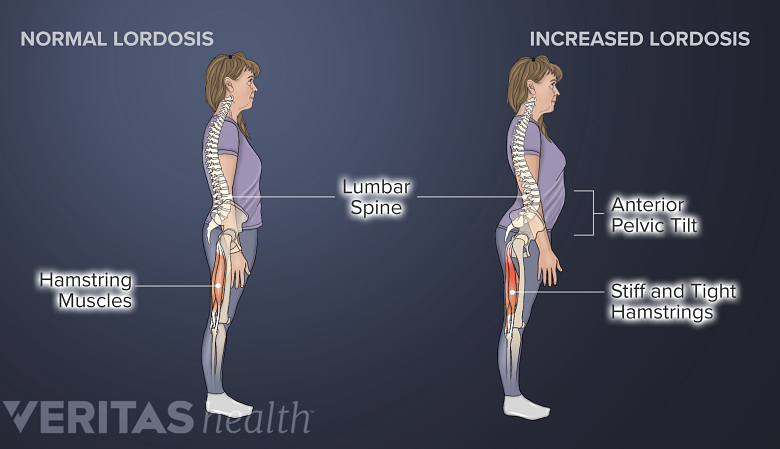
Postural and traumatic changes may cause the lordotic curve to increase, disturbing the balance in the pelvis and hamstring muscles.
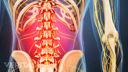
Your lower back (lumbar spine) is the anatomic region between your lowest rib and the upper part of the buttock. 1 Casser HR, Seddigh S, Rauschmann M. Acute Lumbar Back Pain. Dtsch Arztebl Int. 2016;113(13):223–234. doi:10.3238/arztebl.2016.0223 Your spine in this region has a natural inward curve. This curve, called lordosis, helps to:
- Balance the weight of your head on top of your spine
- Evenly distribute weights from your upper body into the lower extremities
- Reduce the concentration of stresses in the lower spine
A problem in your lower back may cause an increase or decrease in this lordosis and may contribute to lower back pain. 2 Cramer GD. The Lumbar Region. In: Clinical Anatomy of the Spine, Spinal Cord, and Ans. Elsevier; 2014:246-311. doi:10.1016/b978-0-323-07954-9.00007-4
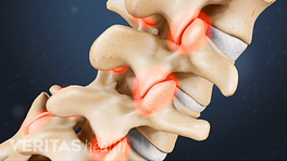
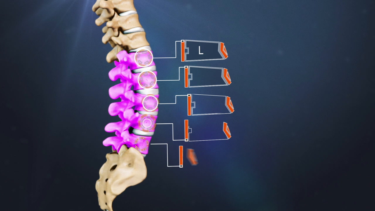
Bones, discs, and joints in your lower back
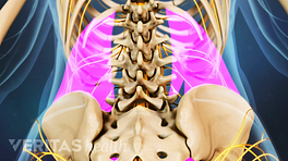
Your lower back contains 5 vertebral bones stacked above each other with intervertebral discs in between. These bones are connected at the back with specialized joints. The lumbar spine connects to the thoracic spine above and the hips below.
Individual anatomical structures include 2 Cramer GD. The Lumbar Region. In: Clinical Anatomy of the Spine, Spinal Cord, and Ans. Elsevier; 2014:246-311. doi:10.1016/b978-0-323-07954-9.00007-4 :
- Vertebrae. Your lumbar vertebrae are labeled L1 to L5, which progressively increase in size, allowing them to bear the body’s weight more effectively. Your vertebrae protect important nervous tissues, such as your spinal cord and the cauda equina.
- Discs. A total of 5 intervertebral discs are situated between your vertebral bodies. The discs typically provide cushioning and shock-absorbing functions to protect your vertebrae during spinal movements.
See Spinal Discs
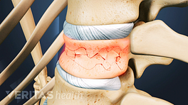
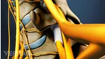
- Facet joints. Your vertebrae are connected in the back of the spine with paired facet joints. These joints provide stability and allow your spine to move in different directions. The joint surfaces are lined by cartilage for smooth movements.
- The facets of the upper lumbar vertebrae are similar to the thoracic facet joints and allow more back and forth spinal movements.
- The facets of the lower lumbar spine are more flexible and facilitate side-to-side movements.
See Facet Joint Disorders and Back Pain
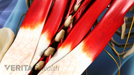
Large muscles and an intricate network of ligaments in your lower back support serve to stabilize your spine and power your twisting and bending movements.
See Back Muscles and Low Back Pain
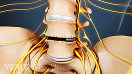
Nerves in your lower back
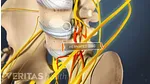
Five pairs of lumbar spinal nerves labeled L1 to L5 branch off your spinal cord and exit through small holes between the vertebrae. The part of the nerve that emerges out of the spine is called the nerve root.
Your lumbar spinal nerves travel down each leg and are formed by 2 types of fibers—sensory fibers that send messages to the brain (when you feel pain after hitting your knee or toe) and motor fibers that receive messages from the brain (when you need to lift your leg to get out of a car or into a bus).
Your lumbar nerves progressively increase in size and contribute to the following functions 4 Dulebohn SC, Ngnitewe Massa R, Mesfin FB. Disc Herniation. [Updated 2019 Aug 1]. In: StatPearls [Internet]. Treasure Island (FL): StatPearls Publishing; 2019 Jan-. Available from: https://www.ncbi.nlm.nih.gov/books/NBK441822/ :
- L1 spinal nerve provides sensation to your groin and genital regions and may contribute to the movement of your hip muscles.
- L2, L3, and L4 spinal nerves provide sensation to the front part of your thigh and along the inner side of your lower leg. These nerves also control movements of your hip and knee muscles.
- L5 spinal nerve provides sensation to the outer side of your lower leg, the upper part of your foot, and the web-space between your first and second toes. Your L5 nerve also controls your hip, knee, foot, and toe movements.
The L4 and L5 nerves (along with other nerves) contribute to the formation of the largest nerve in your body, the sciatic nerve, which runs down from your rear pelvis, into the back of your leg, and terminates in your foot. 5 Giuffre BA, Jeanmonod R. Anatomy, Sciatic Nerve. [Updated 2018 Dec 16]. In: StatPearls [Internet]. Treasure Island (FL): StatPearls Publishing; 2019 Jan-. Available from: https://www.ncbi.nlm.nih.gov/books/NBK482431/. , 6 Davis D, Vasudevan A. Sciatica. [Updated 2019 Feb 28]. In: StatPearls [Internet]. Treasure Island (FL): StatPearls Publishing; 2019 Jan-. Available from: https://www.ncbi.nlm.nih.gov/books/NBK507908/.
Spinal cord and cauda equina in your lower back
Your spinal cord originates in your brain, travels through your spine, and terminates in the upper region of your lower back. This point of termination is called the conus medullaris, 7 Nene Y, Jilani TN. Neuroanatomy, Conus Medullaris. [Updated 2019 Aug 3]. In: StatPearls [Internet]. Treasure Island (FL): StatPearls Publishing; 2019 Jan-. Available from: https://www.ncbi.nlm.nih.gov/books/NBK545227/ from where the spinal nerves descend down. These descending spinal nerves resemble a horse’s tail and are called the cauda equina. 8 Berg EJ, Ashurst JV. Anatomy, Back, Cauda Equina. [Updated 2018 Dec 6]. In: StatPearls [Internet]. Treasure Island (FL): StatPearls Publishing; 2019 Jan-. Available from: https://www.ncbi.nlm.nih.gov/books/NBK513251/
See Spinal Cord and Spinal Nerve Roots
Your spinal cord, conus medullaris, and cauda equina are vital tissues and if they get compressed or damaged, immediate medical attention must be sought.
A basic understanding of the anatomy of your lower back can help you identify and differentiate a problem that commonly affects this region, such as localized muscle pain or sciatica. Knowledge of the structures in your lumbar spine can also help you communicate with your doctor about lower back problems.
Learn More:- 1 Casser HR, Seddigh S, Rauschmann M. Acute Lumbar Back Pain. Dtsch Arztebl Int. 2016;113(13):223–234. doi:10.3238/arztebl.2016.0223
- 2 Cramer GD. The Lumbar Region. In: Clinical Anatomy of the Spine, Spinal Cord, and Ans. Elsevier; 2014:246-311. doi:10.1016/b978-0-323-07954-9.00007-4
- 4 Dulebohn SC, Ngnitewe Massa R, Mesfin FB. Disc Herniation. [Updated 2019 Aug 1]. In: StatPearls [Internet]. Treasure Island (FL): StatPearls Publishing; 2019 Jan-. Available from: https://www.ncbi.nlm.nih.gov/books/NBK441822/
- 5 Giuffre BA, Jeanmonod R. Anatomy, Sciatic Nerve. [Updated 2018 Dec 16]. In: StatPearls [Internet]. Treasure Island (FL): StatPearls Publishing; 2019 Jan-. Available from: https://www.ncbi.nlm.nih.gov/books/NBK482431/.
- 6 Davis D, Vasudevan A. Sciatica. [Updated 2019 Feb 28]. In: StatPearls [Internet]. Treasure Island (FL): StatPearls Publishing; 2019 Jan-. Available from: https://www.ncbi.nlm.nih.gov/books/NBK507908/.
- 7 Nene Y, Jilani TN. Neuroanatomy, Conus Medullaris. [Updated 2019 Aug 3]. In: StatPearls [Internet]. Treasure Island (FL): StatPearls Publishing; 2019 Jan-. Available from: https://www.ncbi.nlm.nih.gov/books/NBK545227/
- 8 Berg EJ, Ashurst JV. Anatomy, Back, Cauda Equina. [Updated 2018 Dec 6]. In: StatPearls [Internet]. Treasure Island (FL): StatPearls Publishing; 2019 Jan-. Available from: https://www.ncbi.nlm.nih.gov/books/NBK513251/