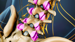
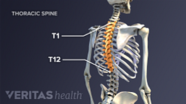
The thoracic spine is comprised of 12 vertebrae labeled T1 through T12. The top thoracic vertebra, T1, connects with C7 in the cervical spine above while the bottom thoracic vertebra, T12, connects with L1 in the lumbar spine below. In addition to being connected to adjacent vertebrae, the thoracic vertebrae are also connected to ribs.
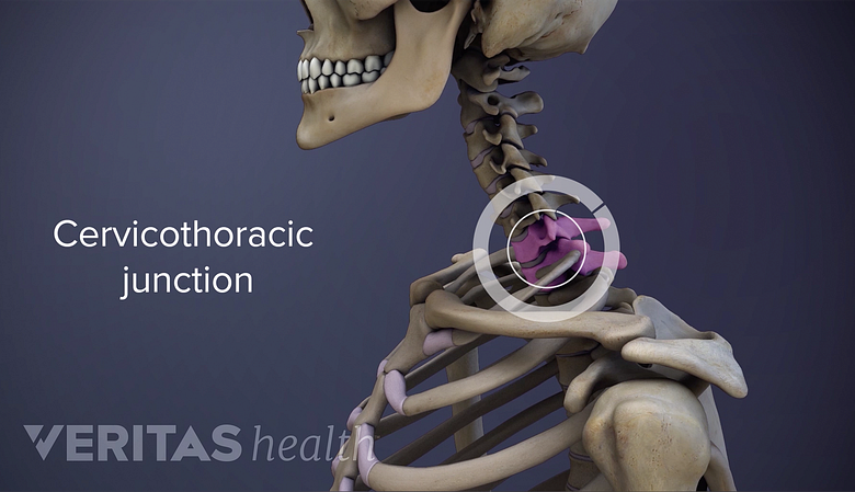
The top of the ribcage begins just below the cervicothoracic junction.
In This Article:
- Thoracic Spine Anatomy and Upper Back Pain
- Thoracic Vertebrae and the Rib Cage
- Thoracic Discs
- Thoracic Spinal Nerves
- Causes of Upper Back Pain Video
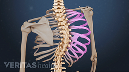
As viewed from the side, the thoracic spine’s vertebrae form a kyphotic curve that runs from T1 to T12, in which the spine curves outward towards the back of the body to allow more room for the internal organs such as the heart and lungs that reside inside the rib cage. Conversely, the cervical spine above and lumbar spine below both have lordotic curves that go inward, toward the front of the body.
Typical Thoracic Vertebrae: T2 to T8
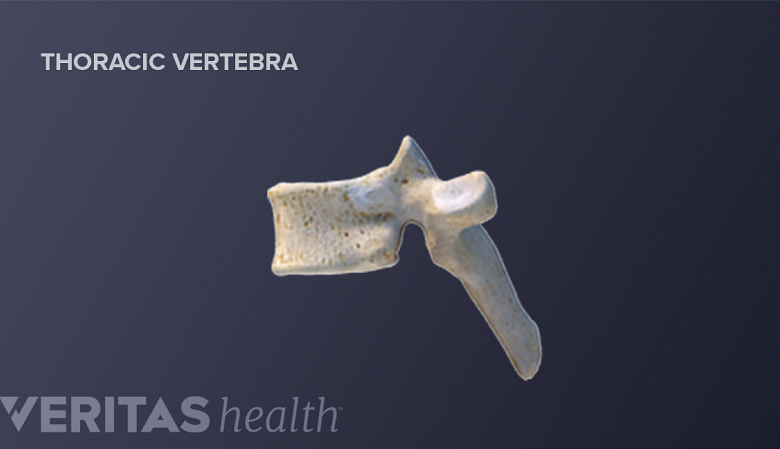
Thoracic vertebrae T2 through T8 all share a similar shape.
Thoracic vertebrae T2 to T8 are all similar, although they do gradually get bigger while going down the spine. A typical thoracic vertebra consists of the following:
- Vertebral body. This thick, bony front of the vertebra is a rounded heart shape (as viewed from above) in the thoracic spine region. The vertebral bodies stack on top of each other and handle most of the spine’s stresses and loads while being cushioned by the intervertebral discs in between.
- Vertebral arch. The vertebral arch forms much of the spinal canal at the sides and back of the vertebrae that encases and protects the spinal cord. (The vertebral body is at the front of the spinal canal.)
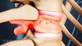
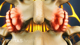
- Facet joints. On each side of the vertebral arch toward the back is one superior (above) and one inferior (below) facet lined with smooth cartilage, which articulate with adjacent vertebrae to form facet joints. These joints are also known as zygapophyseal joints or Z joints.
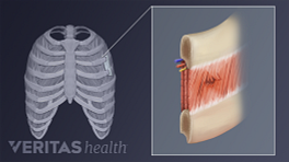
- Costovertebral joints. These joints are where a vertebra connects, or articulates, with a rib. There are two types of costovertebral joints:
- The costocorporeal joint is where the rib head connects with two adjacent vertebral bodies and the disc between them. The bottom of the vertebral body above has a costal demifacet (partial facet), and so does the top of the vertebral body below. These two costal demifacets combine to form a full costal facet for the rib head to articulate between the vertebrae.
- The costotransverse joint is where a small notch near the head of the rib (the tubercle) connects with the vertebra’s lateral extension (the transverse process).
While T2 through T8 are commonly considered typical thoracic vertebrae, there can be variations from person to person regarding which vertebrae are typical and which are unique.
Unique Thoracic Vertebrae
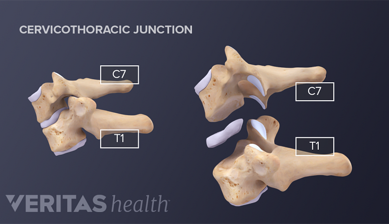
The T1 vertebra has more in common with the cervical vertebrae compared to the other thoracic vertebrae.
The following thoracic vertebrae have unique characteristics that distinguish them from the rest of the thoracic spine:
- T1 is part of the transition from the neck to the upper back, so it shares similar characteristics with the cervical spine compared to the rest of the thoracic spine. Its vertebral body is more rectangular-shaped (not heart-shaped) and lipped (uncinate process up top on each side). The first rib completely articulates with T1 rather than two vertebrae, because T1’s vertebral body has a full upper facet rather than a demifacet.
Watch Spinal Motion Segment: C7-T1 (Cervicothoracic Junction) Animation
- T9 is considered a unique thoracic vertebra if its vertebral body lacks the bottom costal demifacet. In such cases, the T9 vertebral body only articulates with one rib instead of two (the ninth rib but not the tenth).
- T10 does not have an inferior costal demifacet, so its vertebral body only articulates with one rib (the tenth). In some cases, T10’s transverse process does not have a facet to articulate with a rib.
- T11 only has one facet on each side of the vertebra for articulating with the rib head. Also, the transverse process lacks a facet to articulate with a rib.
- T12 also only has one facet on each side of the vertebra. As part of the transition to the lower back, T12 tends to have some characteristics that are more similar to the lumbar spine, such as a larger, wider-shaped vertebral body and a smaller transverse process that is accompanied by two other processes (one that points upward and another that points downward).
How Thoracic Vertebrae Can Contribute to Pain
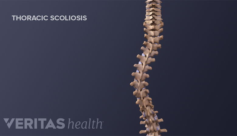
A severe thoracic scoliosis curve may cause the back muscles to become more prone to painful spasms.
A thoracic vertebra can contribute to upper back pain in different ways, such as:
- Trauma. An accident or collision could cause a rib or vertebra to break (fracture) or become misaligned or displaced.
- Osteoarthritis. Wear and tear within the facet joints and/or costovertebral joints can lead to the breakdown of cartilage, resulting in inflammation, osteophytes (bone spurs), and pain.
- Osteoporosis. Bones can weaken with age, especially in women. For people who have osteoporosis, the thoracic vertebrae are the most likely to develop compression fractures that can become painful.
- Kyphosis. The thoracic spine can become hunched forward too much, called kyphosis. The more extreme the kyphosis, the more likely that it can stress muscles to cause upper back pain.
- Scoliosis. Abnormal side-to-side spinal curvature is another possible cause of pain in the upper back if the case is severe.
Thoracic vertebrae can become painful in other ways, such as from infection or other forms of arthritis.