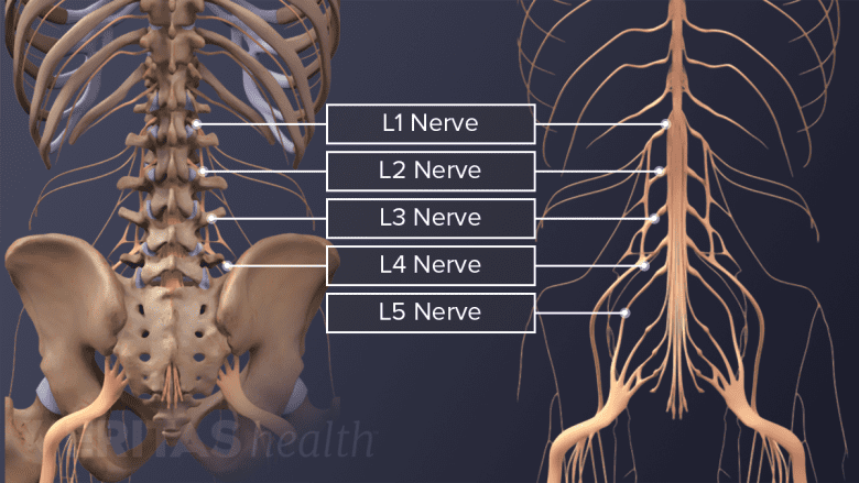
Two spinal nerves branch off from the right and left sides of the spinal cord or the cauda equina at each spinal segment. These spinal nerves are formed by 2 types of fibers—sensory fibers that send messages to the brain (feeling pain when the leg is hurt) and motor fibers that receive messages from the brain (lifting the leg to get out of a car).
There are 5 pairs of lumbar spinal nerves that progressively increase in size from L1 to L5. These nerves exit the intervertebral foramina below the corresponding vertebra. For example, the L4 nerve exits beneath the L4 vertebra through the L4-L5 foramen. These nerves course down from the lower back and merge with other nerves to form the lumbar and lumbosacral plexuses (a network of nerves), which innervate the lower limbs.

The lumbar spinal nerves innervate the lower limbs.
In This Article:
Nerve Root and Spinal Nerve

Nerve roots branch off from both sides of the spinal cord.
The structure of a spinal nerve as it leaves the spinal cord or cauda equina includes:
- Nerve root: Part of the nerve that branches off the spinal cord or cauda equina. At each level, a pair of nerve roots emerge from the right and left sides of the spinal cord. Each pair consists of a dorsal root at the back and a ventral root in the front.
- Spinal nerve: A single nerve formed when the dorsal and ventral nerve roots merge, typically inside the intervertebral foramen (bony opening in between adjacent vertebrae). The spinal nerve travels a short distance inside the intervertebral foramen, after which it branches off into several nerves that innervate different parts of the body.
Doctors may sometimes refer to the part of the spinal nerve exiting the intervertebral foramen as the nerve root or use the terms nerve root and spinal nerve interchangeably.
Dermatomes and Myotomes

Lumbar spinal nerves carry sensory and motor information to the lower body.
The spinal nerves innervate the skin and the muscles of a specific region.
- A dermatome is a specific area of skin that is supplied by the dorsal root fibers of a spinal nerve. The dorsal root fibers carry sensory information from the dermatome to the brain.
- A myotome is a group of muscles controlled by the ventral root fibers of a spinal nerve. The ventral root fibers carry motor signals from the brain to the myotome.
When a spinal nerve gets irritated or compressed, sensory and/or motor deficits may occur in the corresponding dermatome and myotome.
Watch Spinal Cord and Spinal Nerves: What is the Difference? Animation
Functions of the Lumbar Spinal Nerves
The lumbar spinal nerves control movements of the hip and knee muscles.
The 5 pairs of lumbar spinal nerves innervate the lower limbs.
While innervation can vary among individuals, some common patterns include2Dulebohn SC, Ngnitewe Massa R, Mesfin FB. Disc Herniation. [Updated 2019 Aug 1]. In: StatPearls [Internet]. Treasure Island (FL): StatPearls Publishing; 2019 Jan-. Available from: https://www.ncbi.nlm.nih.gov/books/NBK441822/:
- L1 spinal nerve provides sensation to the groin and genital regions and may contribute to the movement of the hip muscles.
- L2, L3, and L4 spinal nerves provide sensation to the front part of the thigh and inner side of the lower leg. These nerves also control movements of the hip and knee muscles.
- L5 spinal nerve provides sensation to the outer side of the lower leg, the upper part of the foot, and the web-space between the first and second toe. The L5 spinal nerve controls hip, knee, foot, and toe movements.
Read more about Spinal Cord and Spinal Nerve Roots
The L4 and L5 nerves (along with other sacral nerves) contribute to the formation of the large sciatic nerve that runs down from the rear pelvis into the back of the leg and terminates in the foot. Symptoms and signs arising from these nerves, typically referred to as sciatica, can cause a sharp, burning pain radiating down the leg with associated weakness and numbness.
While the lumbar spinal nerves progressively increase in size, the openings for these nerves (intervertebral foramina) decrease in size from L1 to L5.1Waxenbaum JA, Futterman B. Anatomy, Back, Lumbar Vertebrae. [Updated 2018 Dec 13]. In: StatPearls [Internet]. Treasure Island (FL): StatPearls Publishing; 2019 Jan-. Available from: https://www.ncbi.nlm.nih.gov/books/NBK459278/ This anatomy, in addition to lower back disorders, such as disc herniation or degeneration may cause the nerve to get compressed, resulting in leg pain and weakness.
- 2 Dulebohn SC, Ngnitewe Massa R, Mesfin FB. Disc Herniation. [Updated 2019 Aug 1]. In: StatPearls [Internet]. Treasure Island (FL): StatPearls Publishing; 2019 Jan-. Available from: https://www.ncbi.nlm.nih.gov/books/NBK441822/
- 1 Waxenbaum JA, Futterman B. Anatomy, Back, Lumbar Vertebrae. [Updated 2018 Dec 13]. In: StatPearls [Internet]. Treasure Island (FL): StatPearls Publishing; 2019 Jan-. Available from: https://www.ncbi.nlm.nih.gov/books/NBK459278/

