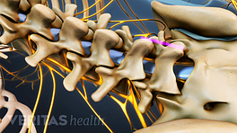
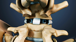
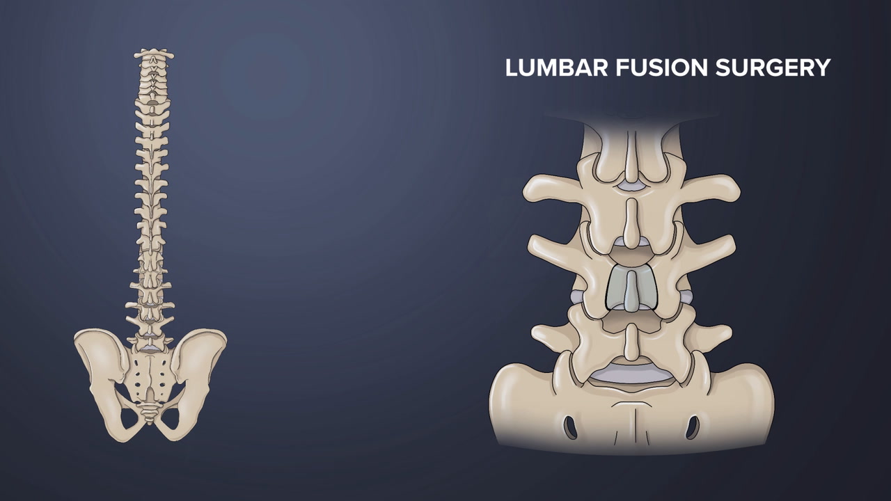
A bone graft is used in spinal fusion surgery, also known as spinal arthrodesis, where additional bone or bone-like tissue is added between the vertebrae to set up the required conditions for the formation of a solid bone bridge.
The solid bone offers stability to the motion segment, protecting neurological structures and providing rigidity to the affected vertebral segments. This rigidity supports weight-bearing functions, such as standing and walking.
A bone graft in spinal fusion surgery sets conditions for solid bone bridge formation.
The fusion does not take place immediately during the surgery; rather, the bone graft placed in between the vertebrae enhances the growth of new bone during the months following the fusion surgery.
In This Article:
- Bone Graft for Spine Fusion
- Autograft: The Patient's Own Bone
- Allograft: Bone Graft from a Donor
- Bone Graft Substitutes
Where Spinal Bone Grafts are Placed
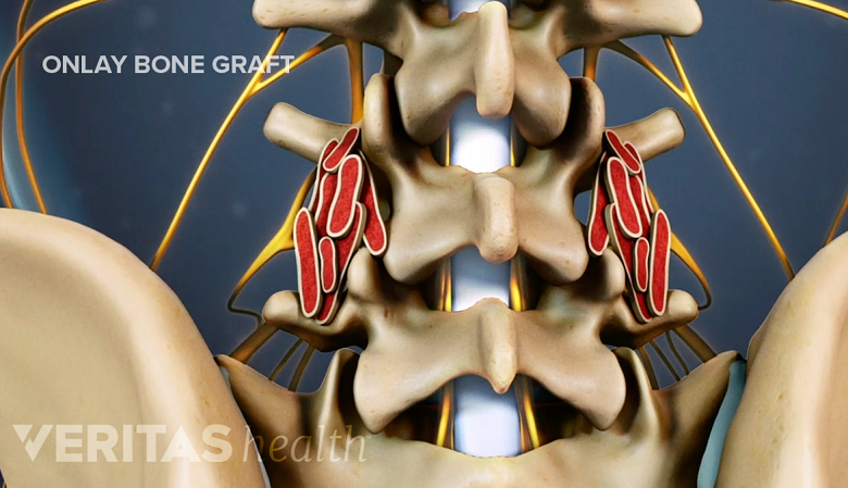
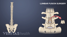
When spinal fusion is approached from the back of the spine, the onlay graft method is used.
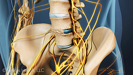
In spinal fusion surgery approached from the front, bone grafts are placed inside implants, such as interbody cages, that are inserted into the space between two vertebrae. Examples of such surgeries include anterior lumbar interbody fusion (ALIF) and anterior cervical discectomy and fusion (ACDF).
In fusion surgeries approached from the back, such as posterior lumbar interbody fusion (PLIF), the bone graft material is placed in the lateral gutter over the facet joints and transverse processes, a method called “onlay graft.” Spinal instrumentation, such as pedicle screws and rods, helps secure the vertebrae together while the onlay bone graft fuses to the facet joints and transverse processes to fuse the two vertebrae together.
Read more about Spine Fusion Instrumentation
Anterior and posterior fusions are sometimes done together to provide better stability and increase fusion rates.
How Spinal Bone Grafts Work
A bone graft consists of living cells and/or fundamental proteins and elements that help commence the fusion process. The graft also provides a scaffold or framework that engages with the blood clot formed at the fusion site to stimulate the growth of new bone and blood vessels. 1 Vaccaro AR, Chiba K, Heller JG, et al. Bone grafting alternatives in spinal surgery. Spine J. 2002;2(3):206-215. doi:10.1016/s1529-9430(02)00180-8 , 2 Orthoinfo by American Academy of Orthopaedic Surgeons. Bone grafts in spine surgery. https://orthoinfo.aaos.org/en/treatment/bone-grafts-in-spine-surgery/. Accessed 09 Aug 2023.
In the United States alone, over half a million bone graft procedures are conducted every year, positioning it as the second most frequently transplanted tissue after blood. 3 Baldwin P, Li DJ, Auston DA, Mir HS, Yoon RS, Koval KJ. Autograft, Allograft, and Bone Graft Substitutes: Clinical Evidence and Indications for Use in the Setting of Orthopaedic Trauma Surgery. J Orthop Trauma. 2019 Apr;33(4):203-213. doi: 10.1097/BOT.0000000000001420. PMID: 30633080.
A bone graft lays the foundation for bone repair and regeneration by facilitating 3 important steps in the fusion process 4 D'Souza M, Macdonald NA, Gendreau JL, Duddleston PJ, Feng AY, Ho AL. Graft Materials and Biologics for Spinal Interbody Fusion. Biomedicines. 2019 Sep 26;7(4):75. doi: 10.3390/biomedicines7040075. PMID: 31561556; PMCID: PMC6966429. :
- Bone resorption: Osteoclast cells are initially responsible for bone resorption (breakdown of bone tissue) upon contact, leading to the formation of tiny cavities on the bone’s surface.
- Bone formation: Next, osteoblast cells occupy these cavities and deposit fresh layers of new bone.
- Bone remodeling: Over time, the entire graft bone or tissue undergoes resorption and replacement, as new bone is continuously laid down, resulting in a complete transformation of the graft.
The type of graft and the presence of a suitable, stabilized environment determine the success of these steps. 4 D'Souza M, Macdonald NA, Gendreau JL, Duddleston PJ, Feng AY, Ho AL. Graft Materials and Biologics for Spinal Interbody Fusion. Biomedicines. 2019 Sep 26;7(4):75. doi: 10.3390/biomedicines7040075. PMID: 31561556; PMCID: PMC6966429.
Characteristics of an Ideal Bone Graft
An ideal bone graft exhibits 3 essential properties 5 Morris MT, Tarpada SP, Cho W. Bone graft materials for posterolateral fusion made simple: a systematic review. Eur Spine J. 2018;27(8):1856-1867. :
- Osteogenic property, which refers to the presence of specialized cells that have the ability to form new bone.
- Osteoinductive property, which refers to the presence of growth factors and chemicals that signal and excite the specialized cells to begin the bone-forming process.
- Osteoconductive property, which refers to a structural scaffolding where the bone-forming cells can lay down new bone.
The iliac crest (hip bone) satisfies all the above criteria and is commonly used as a graft material in spinal fusion. 5 Morris MT, Tarpada SP, Cho W. Bone graft materials for posterolateral fusion made simple: a systematic review. Eur Spine J. 2018;27(8):1856-1867.
How Long Does the Bone Healing Process Take for Spinal Fusion
While the bone fusion timeline is variable, as a general guideline, the initial phase of the bone graft healing process takes approximately 1 week to 1 month following the surgery, 6 Sheen JR, Garla VV. Fracture Healing Overview. [Updated 2022 May 8]. In: StatPearls [Internet]. Treasure Island (FL): StatPearls Publishing; 2023 Jan-. Available from: https://www.ncbi.nlm.nih.gov/books/NBK551678 , 7 Cruz A, Ropper AE, Xu DS, et al. Failure in Lumbar Spinal Fusion and Current Management Modalities. Semin Plast Surg. 2021;35(1):54-62. doi:10.1055/s-0041-1726102 and certain activity restrictions continue for up to 6 months or longer. For the solid bone to completely heal, it can take up to several months to years. 6 Sheen JR, Garla VV. Fracture Healing Overview. [Updated 2022 May 8]. In: StatPearls [Internet]. Treasure Island (FL): StatPearls Publishing; 2023 Jan-. Available from: https://www.ncbi.nlm.nih.gov/books/NBK551678 , 7 Cruz A, Ropper AE, Xu DS, et al. Failure in Lumbar Spinal Fusion and Current Management Modalities. Semin Plast Surg. 2021;35(1):54-62. doi:10.1055/s-0041-1726102 Load-bearing activities provide stimulation to the fusion and enhance the strength of the fused bone. 7 Cruz A, Ropper AE, Xu DS, et al. Failure in Lumbar Spinal Fusion and Current Management Modalities. Semin Plast Surg. 2021;35(1):54-62. doi:10.1055/s-0041-1726102
Though the process of fusion and remodeling occurs over an extended period of time, most patients can resume activities of daily living, including weight-bearing activities, such as returning to work and exercising over a period of a few weeks. 8 Guglielmi GN, Seibly JM. Return to Work Guidelines Following Neurosurgical Procedures. Cureus. 2020;12(12):e11982. Published 2020 Dec 8. doi:10.7759/cureus.11982
Bone healing takes place in three stages, categorized by the type of cellular activity 4 D'Souza M, Macdonald NA, Gendreau JL, Duddleston PJ, Feng AY, Ho AL. Graft Materials and Biologics for Spinal Interbody Fusion. Biomedicines. 2019 Sep 26;7(4):75. doi: 10.3390/biomedicines7040075. PMID: 31561556; PMCID: PMC6966429. :
| Healing stage | Duration | Cellular Activity |
|---|---|---|
| Inflammatory stage | The inflammatory stage lasts for hours to days post-surgery. | In this stage, there’s an infiltration of cells that fight inflammation, along with cells that help build new bone, such as osteoprogenitor cells and fibroblasts. |
| Repair stage | The repair stage spans several weeks or months after surgery. | In this stage, new blood vessels, bone tissue, and structures or matrices made of collagen are formed at the site of bone fusion. The gap between the vertebrae begins to close and a bridge is formed. |
| Bone remodeling stage | The bone remodeling stage lasts for months to years after surgery. | This stage focuses on gradually reshaping and strengthening the newly laid bone tissue through mechanical stress placed on the spine. |
Within these stages, the body’s biological bone healing process is complex, influenced by the cells, chemical processes, and mechanical stresses on the spinal segment.
The bone fusion process is variable and depends on a number of factors, which is why surgeons follow up with patients and monitor their individual postoperative status.
Watch Video: Stages of Bone Healing in Lumbar Spine Fusion Surgery
5 Types of Bone Graft Materials Used in Spinal Fusion
Bone grafts are broadly categorized depending on their source. Each type of graft works differently and has its own set of potential benefits and risks.
1. Real bone tissue
Real bone tissue is harvested from the patient or obtained from a donor and serves as a vital resource for spinal grafting. Examples include 4 D'Souza M, Macdonald NA, Gendreau JL, Duddleston PJ, Feng AY, Ho AL. Graft Materials and Biologics for Spinal Interbody Fusion. Biomedicines. 2019 Sep 26;7(4):75. doi: 10.3390/biomedicines7040075. PMID: 31561556; PMCID: PMC6966429. :
- Autograft, which is the patient’s own bone. This bone is taken from the hip or another part of the body at the same time the fusion surgery is performed. Autografts offer a variety of potential benefits and are regarded as the gold standard for spinal fusion grafting.
- Allografts, which are obtained from a deceased or living donor (an example of a living donor is someone who recently underwent hip replacement surgery).
- Demineralized bone matrices, which are organic matrices of allograft bone tissue where the mineralized part of the bone is removed using acid. The organic matrix is composed of collagen-based and non-collagenous proteins, along with growth factors.
Real bone grafts have demonstrated high success rates in promoting successful spinal fusion outcomes, 10 Wind J, Park D, Lansford T, Nunley P, Peppers T, Russo A, Hassanzadeh H, Sembrano JN, Yoo J, Sales J. Twelve-Month Results from a Prospective Clinical Study Evaluating the Efficacy and Safety of Cellular Bone Allograft in Subjects Undergoing Lumbar Spinal Fusion. Neurol Int. 2022 Oct 26;14(4):875-883. doi: 10.3390/neurolint14040070. PMID: 36412692; PMCID: PMC9680433. , 11 Arnold PM, Sasso RC, Janssen ME, Fehlings MG, Smucker JD, Vaccaro AR, Heary RF, Patel AI, Goulet B, Kalfas IH, Kopjar B. Efficacy of i-Factor Bone Graft versus Autograft in Anterior Cervical Discectomy and Fusion: Results of the Prospective, Randomized, Single-blinded Food and Drug Administration Investigational Device Exemption Study. Spine (Phila Pa 1976). 2016 Jul 1;41(13):1075-1083. doi: 10.1097/BRS.0000000000001466. PMID: 26825787. especially in challenging cases or patients with compromised bone health.
2. Bone tissue substitutes
Bone substitutes are synthetic or biologically derived materials that mimic the properties of natural bone and promote bone regeneration and fusion. Examples include 4 D'Souza M, Macdonald NA, Gendreau JL, Duddleston PJ, Feng AY, Ho AL. Graft Materials and Biologics for Spinal Interbody Fusion. Biomedicines. 2019 Sep 26;7(4):75. doi: 10.3390/biomedicines7040075. PMID: 31561556; PMCID: PMC6966429. :
- Ceramics, which are calcium-based compounds that have a similar structure and mineral composition as natural bone.
- Bone morphogenic proteins, which contain cytokines (substances secreted by the cells of the immune system) and growth factors that help initiate bone healing.
Bone graft substitutes are readily available, eliminating the need for harvesting bone from another part of the patient's body.
3. Autologous growth factors
Autologous growth factors are special substances extracted from the platelets (cells in the blood), which have the ability to induce bone growth. Two of these substances – platelet-derived growth factor (PDGF) and TGF-β are particularly studied in spinal fusion. PDGF promotes remodeling and construction of new bone, while TGF-β provides a scaffolding for fusion. 4 D'Souza M, Macdonald NA, Gendreau JL, Duddleston PJ, Feng AY, Ho AL. Graft Materials and Biologics for Spinal Interbody Fusion. Biomedicines. 2019 Sep 26;7(4):75. doi: 10.3390/biomedicines7040075. PMID: 31561556; PMCID: PMC6966429.
Autologous growth factors help enhance the effectiveness of bone fusion processes when they are combined with autografts, allografts, or bone substitutes. 4 D'Souza M, Macdonald NA, Gendreau JL, Duddleston PJ, Feng AY, Ho AL. Graft Materials and Biologics for Spinal Interbody Fusion. Biomedicines. 2019 Sep 26;7(4):75. doi: 10.3390/biomedicines7040075. PMID: 31561556; PMCID: PMC6966429.
4. Mesenchymal stem cells
Mesenchymal stem cells have the ability to differentiate into bone and cartilage cells, such as osteoblasts and chondrocytes, making them useful in enabling spinal fusion. These cells are usually harvested from the hip bone (iliac crest) and deposited into the graft bone. 4 D'Souza M, Macdonald NA, Gendreau JL, Duddleston PJ, Feng AY, Ho AL. Graft Materials and Biologics for Spinal Interbody Fusion. Biomedicines. 2019 Sep 26;7(4):75. doi: 10.3390/biomedicines7040075. PMID: 31561556; PMCID: PMC6966429.
5. Synthetic peptides
Man-made peptides, such as P-15 (a peptide with 15 amino acid chains), have the ability to mimic the bone mineralization process when combined with specific calcium-containing granules. Together with these granules, this peptide provides the scaffolding and source of calcium for new bone formation. 4 D'Souza M, Macdonald NA, Gendreau JL, Duddleston PJ, Feng AY, Ho AL. Graft Materials and Biologics for Spinal Interbody Fusion. Biomedicines. 2019 Sep 26;7(4):75. doi: 10.3390/biomedicines7040075. PMID: 31561556; PMCID: PMC6966429.
More research is needed to establish the long-term effects of fusion and rates of successful fusion with this type of graft. 4 D'Souza M, Macdonald NA, Gendreau JL, Duddleston PJ, Feng AY, Ho AL. Graft Materials and Biologics for Spinal Interbody Fusion. Biomedicines. 2019 Sep 26;7(4):75. doi: 10.3390/biomedicines7040075. PMID: 31561556; PMCID: PMC6966429.
Selecting a Bone Graft for Spinal Fusion
Selecting an appropriate bone graft is influenced by factors related to the surgery, patient, and surgeon, and typically include:
- Type of spinal fusion (eg, ALIF, PLIF, or posterolateral gutter fusion)
- The number of levels of the spine involved (single-level fusions vs multi-level spinal fusion)
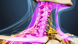
- Location of the fusion – cervical (neck) fusion or lumbar (low back) fusion
- Patient risk factors for potential non-fusion (eg, if the patient is obese, a smoker, or has poor bone quality)
- Surgeon experience and preference
An appropriately selected bone graft helps balance biocompatibility, improve structural support, and stimulate new bone formation in the patient. A tailored choice can enhance fusion and expedite post-surgery recovery.
Factors to Achieve a Solid Fusion
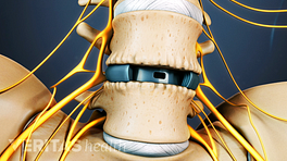
Spinal interbody cages promote bone healing by dissipating forces placed on the treated segment.
A major risk of spinal fusion surgery is the failure to relieve lower back pain symptoms following the surgery. This failed outcome is colloquially referred to as failed back surgery syndrome. 12 Li T, Shi L, Luo Y, Chen D, Chen Y. One-level or multilevel interbody fusion for multilevel lumbar degenerative diseases: a prospective randomized control study with a 4-year follow-up. World Neurosurgery. 2018;110:e815-e822. doi:10.1016/j.wneu.2017.11.109 A common cause of failed back surgery syndrome is pseudoarthrosis, which refers to the failure of the spinal segment to properly heal and fuse into a solid, strong bony segment. 13 Tavares WM, de França SA, Paiva WS, Teixeira MJ. A systematic review and meta-analysis of fusion rate enhancements and bone graft options for spine surgery. Sci Rep. 2022 May 9;12(1):7546. doi: 10.1038/s41598-022-11551-8. PMID: 35534520; PMCID: PMC9085837.
A few examples of factors that influence the formation of a solid fusion from bone graft include:
- Smoking. Smoking releases nicotine into the body, which causes imbalances in the body’s metabolic functions, decreases bone mineral density, reduces bone formation, and ultimately delays bone healing, which may result in failed fusion. 14 Cruz A, Ropper AE, Xu DS, et al. Failure in Lumbar Spinal Fusion and Current Management Modalities. Semin Plast Surg. 2021;35(1):54-62. doi:10.1055/s-0041-1726102
- Extent and type of surgery. Multilevel fusions that involve 3 or more levels have a significantly higher risk of developing pseudoarthrosis. This risk is particularly high in fusions involving the L5-S1 spinal segment. 15 Tannoury C, Bhale R, Vora M, Saade A, Kortbawi R, Orlando G, Das A, Tannoury T. Pseudarthrosis Following Lumbar and Lumbosacral Fusion Using the Antepsoas Technique. Spine (Phila Pa 1976). 2021 Dec 15;46(24):1690-1695. doi: 10.1097/BRS.0000000000004115
- Use of interbody cages. A spinal interbody cage is filled with bone graft material that grows over time and fills within the interbody space. These cages are designed to promote bone healing by dissipating forces placed on the treated segment. 16 Souslian FG, Patel PD. Review and analysis of modern lumbar spinal fusion techniques. Br J Neurosurg. 2021 Jul 15:1-7. doi: 10.1080/02688697.2021.1881041. Epub ahead of print. PMID: 34263676.
The above is by no means a complete list and is provided to illustrate the influence of factors involved in growing new bone from a bone graft.
Read more about Failed Spinal Fusion Surgery
- 1 Vaccaro AR, Chiba K, Heller JG, et al. Bone grafting alternatives in spinal surgery. Spine J. 2002;2(3):206-215. doi:10.1016/s1529-9430(02)00180-8
- 2 Orthoinfo by American Academy of Orthopaedic Surgeons. Bone grafts in spine surgery. https://orthoinfo.aaos.org/en/treatment/bone-grafts-in-spine-surgery/. Accessed 09 Aug 2023.
- 3 Baldwin P, Li DJ, Auston DA, Mir HS, Yoon RS, Koval KJ. Autograft, Allograft, and Bone Graft Substitutes: Clinical Evidence and Indications for Use in the Setting of Orthopaedic Trauma Surgery. J Orthop Trauma. 2019 Apr;33(4):203-213. doi: 10.1097/BOT.0000000000001420. PMID: 30633080.
- 4 D'Souza M, Macdonald NA, Gendreau JL, Duddleston PJ, Feng AY, Ho AL. Graft Materials and Biologics for Spinal Interbody Fusion. Biomedicines. 2019 Sep 26;7(4):75. doi: 10.3390/biomedicines7040075. PMID: 31561556; PMCID: PMC6966429.
- 5 Morris MT, Tarpada SP, Cho W. Bone graft materials for posterolateral fusion made simple: a systematic review. Eur Spine J. 2018;27(8):1856-1867.
- 6 Sheen JR, Garla VV. Fracture Healing Overview. [Updated 2022 May 8]. In: StatPearls [Internet]. Treasure Island (FL): StatPearls Publishing; 2023 Jan-. Available from: https://www.ncbi.nlm.nih.gov/books/NBK551678
- 7 Cruz A, Ropper AE, Xu DS, et al. Failure in Lumbar Spinal Fusion and Current Management Modalities. Semin Plast Surg. 2021;35(1):54-62. doi:10.1055/s-0041-1726102
- 8 Guglielmi GN, Seibly JM. Return to Work Guidelines Following Neurosurgical Procedures. Cureus. 2020;12(12):e11982. Published 2020 Dec 8. doi:10.7759/cureus.11982
- 10 Wind J, Park D, Lansford T, Nunley P, Peppers T, Russo A, Hassanzadeh H, Sembrano JN, Yoo J, Sales J. Twelve-Month Results from a Prospective Clinical Study Evaluating the Efficacy and Safety of Cellular Bone Allograft in Subjects Undergoing Lumbar Spinal Fusion. Neurol Int. 2022 Oct 26;14(4):875-883. doi: 10.3390/neurolint14040070. PMID: 36412692; PMCID: PMC9680433.
- 11 Arnold PM, Sasso RC, Janssen ME, Fehlings MG, Smucker JD, Vaccaro AR, Heary RF, Patel AI, Goulet B, Kalfas IH, Kopjar B. Efficacy of i-Factor Bone Graft versus Autograft in Anterior Cervical Discectomy and Fusion: Results of the Prospective, Randomized, Single-blinded Food and Drug Administration Investigational Device Exemption Study. Spine (Phila Pa 1976). 2016 Jul 1;41(13):1075-1083. doi: 10.1097/BRS.0000000000001466. PMID: 26825787.
- 12 Li T, Shi L, Luo Y, Chen D, Chen Y. One-level or multilevel interbody fusion for multilevel lumbar degenerative diseases: a prospective randomized control study with a 4-year follow-up. World Neurosurgery. 2018;110:e815-e822. doi:10.1016/j.wneu.2017.11.109
- 13 Tavares WM, de França SA, Paiva WS, Teixeira MJ. A systematic review and meta-analysis of fusion rate enhancements and bone graft options for spine surgery. Sci Rep. 2022 May 9;12(1):7546. doi: 10.1038/s41598-022-11551-8. PMID: 35534520; PMCID: PMC9085837.
- 14 Cruz A, Ropper AE, Xu DS, et al. Failure in Lumbar Spinal Fusion and Current Management Modalities. Semin Plast Surg. 2021;35(1):54-62. doi:10.1055/s-0041-1726102
- 15 Tannoury C, Bhale R, Vora M, Saade A, Kortbawi R, Orlando G, Das A, Tannoury T. Pseudarthrosis Following Lumbar and Lumbosacral Fusion Using the Antepsoas Technique. Spine (Phila Pa 1976). 2021 Dec 15;46(24):1690-1695. doi: 10.1097/BRS.0000000000004115
- 16 Souslian FG, Patel PD. Review and analysis of modern lumbar spinal fusion techniques. Br J Neurosurg. 2021 Jul 15:1-7. doi: 10.1080/02688697.2021.1881041. Epub ahead of print. PMID: 34263676.