Magnetic resonance imaging (MRI) is a powerful diagnostic tool used to evaluate spine pathology.
MRI scan results alone are not diagnostic.
After your physician takes a thorough history and performs and physical examination an MRI can help confirm or refine a suspected diagnosis.
If you don’t fit the typical guidelines for an MRI, be sure to read to the end to understand exceptions to the rule.

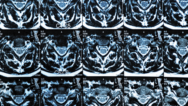
MRI scan of the cervical spine showing multiple spinal segments
When is an MRI Typically Recommended?
An MRI is typically recommended in one of three cases: after 6 weeks of nonsurgical treatment, to plan for minimally invasive procedures or to plan for surgery.
1. 6 Weeks of Non-Surgical Treatments
An MRI is commonly recommended only after at least 6 weeks of non-surgical treatments, such as physical therapy, medications, heat and cold therapy, activity restrictions and relative rest.
If pain and/or related symptoms persist for more than six weeks despite nonoperative treatment, an MRI may be ordered to image the spine.
This is particularly true if there is nerve involvement and your pain radiates into your arms or legs, is accompanied by numbness or weakness, or if neurological symptoms are worsening.
2. Injections and Minimally Invasive Procedure Planning
An MRI is often used to guide treatment decisions for non-operative procedures such as injections.
For example:
- It is common practice to do an MRI scan prior to doing an epidural steroid injection for severe pain from a herniated disc
- An MRI can detect endplate damage which can help determine if a patient is a good candidate for a basivertebral nerve ablation
3. Planning Spine Surgery
An MRI is almost always done prior to spine surgery to inform surgical decisions. For example:
- If a degenerated disc is compressing the spinal cord or nerves, an MRI can help determine whether a surgical approach from the front of the spine (such as an anterior lumbar interbody fusion, or ALIF) or the back of the spine (such as a posterior lumbar interbody fusion, or PLIF) is more appropriate.
- In cases of spinal stenosis, an MRI can show the extent of nerve compression and help the surgeon decide whether a decompression surgery or a spinal fusion is needed.
- For patients with scoliosis or other spinal deformities, an MRI can provide detailed images of the curvature and any associated nerve involvement, aiding surgical planning.
Exceptions to the 6-week minimum guideline
There are specific situations in which an MRI scan may be recommended earlier and without the minimum 6-weeks of nonsurgical treatments, including:
Emergency Situations: An MRI is immediately indicated if there are signs of serious conditions such as:
- Spinal cord compression: Symptoms may include sudden weakness, numbness, or loss of bowel or bladder control.
- Suspected tumors: Symptoms are accompanied by unexplained weight loss, fever, or severe night pain.
- Infections: Symptoms may include fever, chills, night sweats and localized back pain.
Severe Disability or Neurological Issues: An MRI may be performed earlier than 6 weeks if there is severe disability, such as the inability to walk, or if neurological symptoms are progressing rapidly.
For example, if a patient develops worsening foot drop (inability to lift the front part of the foot), an MRI may be necessary to assess nerve compression.
Trauma: An MRI may be recommended sooner if you have concerning symptoms along with a trauma, such as a sports injury or car accident affecting your neck or back.
MRI for Surgical Planning
MRI is most frequently used to plan spine surgeries.
This scan provides detailed images of soft tissues, including discs, nerves, and the spinal cord, which are not visible on X-rays or CT scans.
This information is critical for determining the best surgical approach. For example:
- If a herniated disc is compressing a nerve root, an MRI can help the surgeon decide whether a microdiscectomy or endoscopic approach is appropriate.
- In cases of severe spinal stenosis, an MRI can show the exact location and severity of the narrowing, helping the surgeon plan a laminectomy or other decompression procedure.
Always consult with a spine specialist to determine whether an MRI is appropriate for your specific situation.
Remember, the results of an MRI need to be interpreted in the context of your symptoms, medical history, and physical examination findings to guide the best course of treatment.

